Brunet Sylvain, Scheuer Todd, Klevit Rachel, Catterall William A
Department of Pharmacology, University of Washington, Seattle, WA 98195, USA.
J Gen Physiol. 2005 Oct;126(4):311-23. doi: 10.1085/jgp.200509333. Epub 2005 Sep 12.
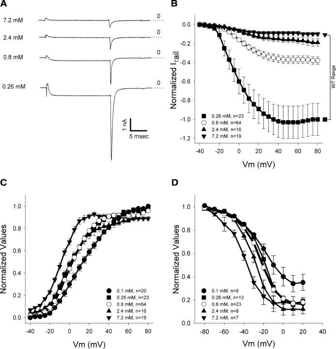
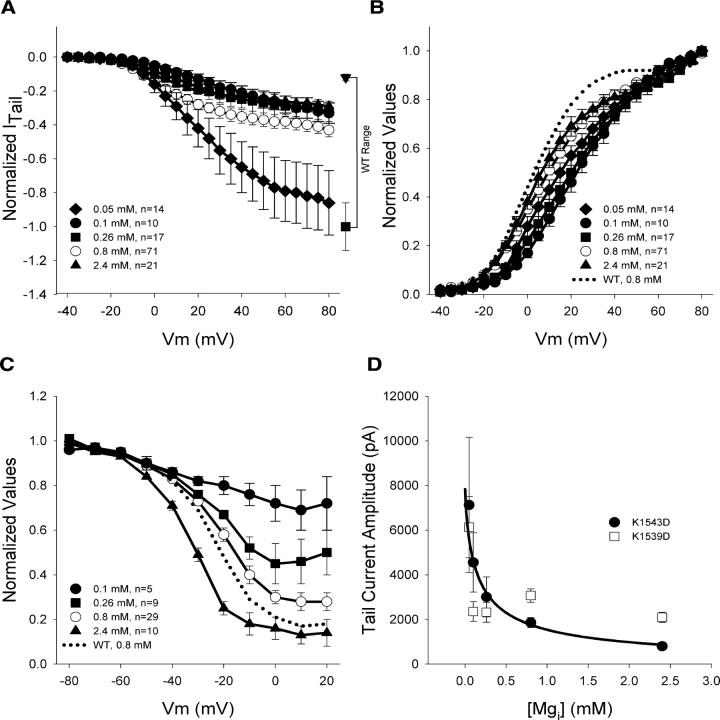
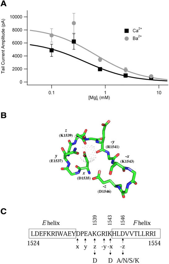
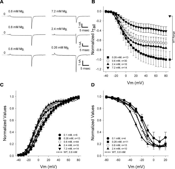
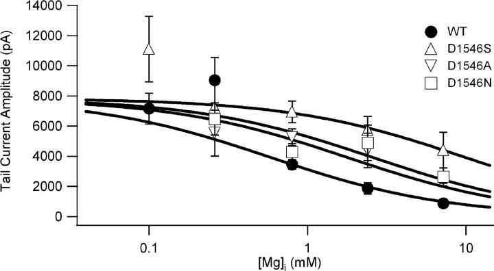
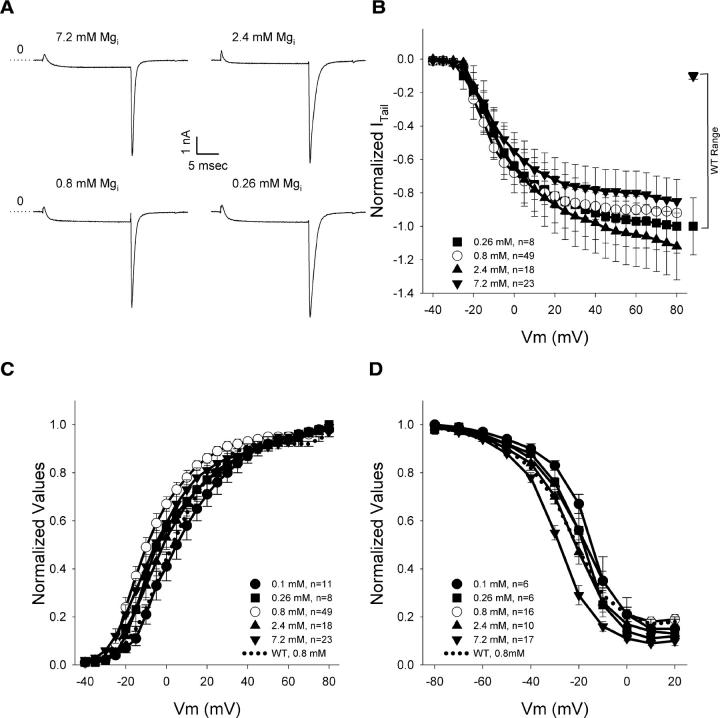
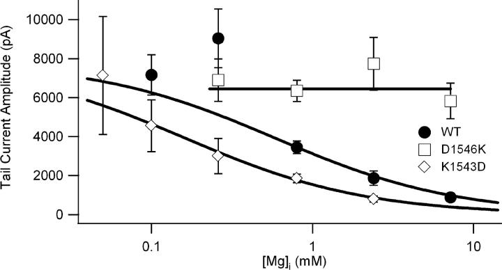
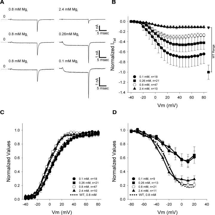
Magnesium levels in cardiac myocytes change in cardiovascular diseases. Intracellular free magnesium (Mg(i)) inhibits L-type Ca(2+) currents through Ca(V)1.2 channels in cardiac myocytes, but the mechanism of this effect is unknown. We hypothesized that Mg(i) acts through the COOH-terminal EF-hand of Ca(V)1.2. EF-hand mutants were engineered to have either decreased (D1546A/N/S/K) or increased (K1543D and K1539D) Mg(2+) affinity. In whole-cell patch clamp experiments, increased Mg(i) reduced both Ba(2+) and Ca(2+) currents conducted by wild type (WT) Ca(V)1.2 channels expressed in tsA-201 cells with similar affinity. Exposure of WT Ca(V)1.2 to lower Mg(i) (0.26 mM) increased the amplitudes of Ba(2+) currents 2.6 +/- 0.4-fold without effects on the voltage dependence of activation and inactivation. In contrast, increasing Mg(i) to 2.4 or 7.2 mM reduced current amplitude to 0.5 +/- 0.1 and 0.26 +/- 0.05 of the control level at 0.8 mM Mg(i). The effects of Mg(i) on peak Ba(2+) currents were approximately fit by a single binding site model with an apparent K(d) of 0.65 mM. The apparent K(d) for this effect of Mg(i) was shifted approximately 3.3- to 16.5-fold to higher concentration in D1546A/N/S mutants, with only small effects on the voltage dependence of activation and inactivation. Moreover, mutant D1546K was insensitive to Mg(i) up to 7.2 mM. In contrast to these results, peak Ba(2+) currents through the K1543D mutant were inhibited by lower concentrations of Mg(i) compared with WT, consistent with approximately fourfold reduction in apparent K(d) for Mg(i), and inhibition of mutant K1539D by Mg(i) was also increased comparably. In addition to these effects, voltage-dependent inactivation of K1543D and K1539D was incomplete at positive membrane potentials when Mg(i) was reduced to 0.26 or 0.1 mM, respectively. These results support a novel mechanism linking the COOH-terminal EF-hand with modulation of Ca(V)1.2 channels by Mg(i). Our findings expand the repertoire of modulatory interactions taking place at the COOH terminus of Ca(V)1.2 channels, and reveal a potentially important role of Mg(i) binding to the COOH-terminal EF-hand in regulating Ca(2+) influx in physiological and pathophysiological states.
心肌细胞中的镁水平在心血管疾病中会发生变化。细胞内游离镁(Mg(i))通过心肌细胞中的Ca(V)1.2通道抑制L型Ca(2+)电流,但其作用机制尚不清楚。我们推测Mg(i)通过Ca(V)1.2的COOH末端EF手结构域发挥作用。构建了EF手突变体,使其对Mg(2+)的亲和力降低(D1546A/N/S/K)或增加(K1543D和K1539D)。在全细胞膜片钳实验中,细胞内镁增加以相似的亲和力降低了tsA-201细胞中野生型(WT)Ca(V)1.2通道传导的Ba(2+)和Ca(2+)电流。将WT Ca(V)1.2暴露于较低的Mg(i)(0.26 mM)中可使Ba(2+)电流幅度增加2.6±0.4倍,而对激活和失活的电压依赖性无影响。相反,将Mg(i)增加到2.4或7.2 mM时,电流幅度在0.8 mM Mg(i)时分别降至对照水平的0.5±0.1和0.26±0.05。Mg(i)对Ba(2+)电流峰值的影响大致符合单结合位点模型,表观解离常数(K(d))为0.65 mM。Mg(i)这种作用的表观K(d)在D1546A/N/S突变体中向更高浓度偏移了约3.3至16.5倍,对激活和失活的电压依赖性影响较小。此外,突变体D1546K在高达7.2 mM的Mg(i)下对其不敏感。与这些结果相反,与WT相比,通过K1543D突变体的Ba(2+)电流峰值受到较低浓度Mg(i)的抑制,这与Mg(i)的表观K(d)降低约四倍一致,并且Mg(i)对突变体K1539D的抑制作用也相应增加。除了这些影响外,当Mg(i)分别降至0.26或0.1 mM时,K1543D和K1539D在正膜电位下的电压依赖性失活不完全。这些结果支持了一种将COOH末端EF手结构域与Mg(i)对Ca(V)1.2通道的调节联系起来的新机制。我们的发现扩展了在Ca(V)1.2通道COOH末端发生的调节相互作用的范围,并揭示了Mg(i)与COOH末端EF手结构域结合在生理和病理生理状态下调节Ca(2+)内流中的潜在重要作用。