Hahn P J, Nevaldine B, Longo J A
Department of Radiology, State University of New York Health Science Center, Syracuse 13210.
Mol Cell Biol. 1992 Jul;12(7):2911-8. doi: 10.1128/mcb.12.7.2911-2918.1992.
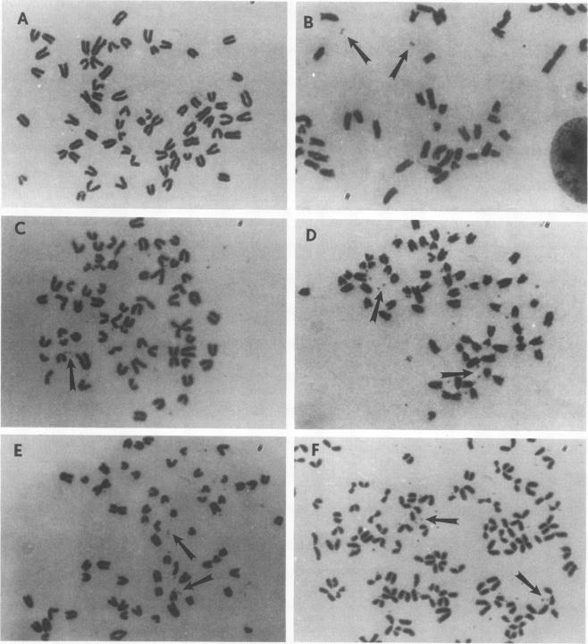
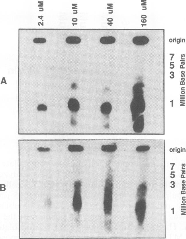
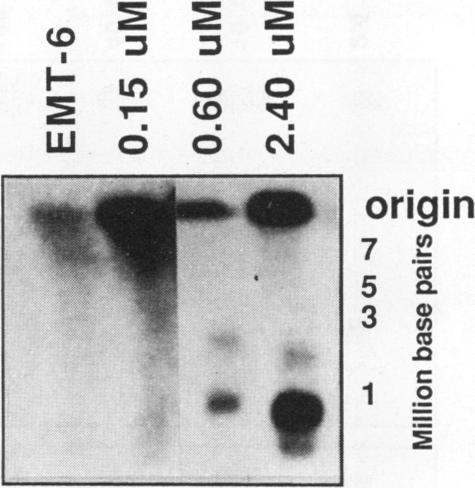
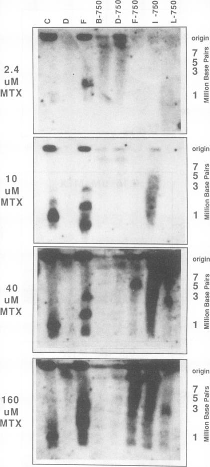
To determine whether microscopically visible double-minute chromosomes (DMs) are derived from submicroscopic precursors, we monitored the amplification of the dihydrofolate reductase (DHFR) gene in 10 independent isolates of methotrexate (MTX)-resistant mouse cells. At every other doubling in MTX concentration, the cells were examined both microscopically, to detect the presence of microscopically visible DMs, and by pulsed-field gel electrophoresis and hybridization to a DHFR-specific probe, to detect submicroscopic DMs. One of the cloned MTX-resistant isolates was examined in detail and was shown to originally contain amplified DHFR genes on circular DMs measuring 1 and 3 Mb in size; additionally, metaphase chromosome preparations from this cloned isolate were examined and were shown to contain microscopically visible DMs too large to enter a pulsed-field gel. During stepwise selection for increasing levels of MTX, the smaller DMs (not microscopically visible) were shown to be preferentially amplified, whereas the larger (microscopically visible) ones decreased in relative numbers. Rare-cutting NotI digestion patterns of total genomic DNA that includes the DMs containing the DHFR gene suggest that the DMs increase in copy number without any further significant rearrangements. We saw no evidence from any of the 10 isolates to suggest that microscopically visible DMs are formed from smaller submicroscopic precursors.
为了确定显微镜下可见的双微小染色体(DMs)是否源自亚显微前体,我们监测了10株独立的耐甲氨蝶呤(MTX)小鼠细胞系中二氢叶酸还原酶(DHFR)基因的扩增情况。在MTX浓度每隔一次加倍时,对细胞进行显微镜检查以检测显微镜下可见的DMs的存在,并通过脉冲场凝胶电泳和与DHFR特异性探针杂交来检测亚显微DMs。对其中一株克隆的耐MTX分离株进行了详细检查,结果显示其最初在大小为1和3 Mb的环状DMs上含有扩增的DHFR基因;此外,对该克隆分离株的中期染色体标本进行检查,发现其中含有显微镜下可见的、太大而无法进入脉冲场凝胶的DMs。在逐步选择更高水平的MTX过程中,较小的DMs(显微镜下不可见)被优先扩增,而较大的(显微镜下可见)DMs相对数量减少。包含含有DHFR基因的DMs的总基因组DNA的稀有切割NotI消化模式表明,DMs的拷贝数增加而没有任何进一步的显著重排。在这10株分离株中,我们没有发现任何证据表明显微镜下可见的DMs是由较小的亚显微前体形成的。