Nomura T, Honmou O, Harada K, Houkin K, Hamada H, Kocsis J D
Department of Neurosurgery, Sapporo Medical University School of Medicine, South-1st, West-16th, Chuo-ku, Sapporo, Hokkaido 060-8543, Japan.
Neuroscience. 2005;136(1):161-9. doi: 10.1016/j.neuroscience.2005.06.062. Epub 2005 Oct 17.
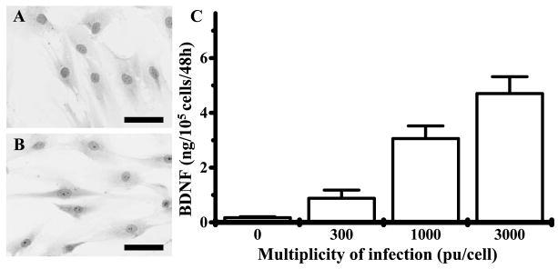
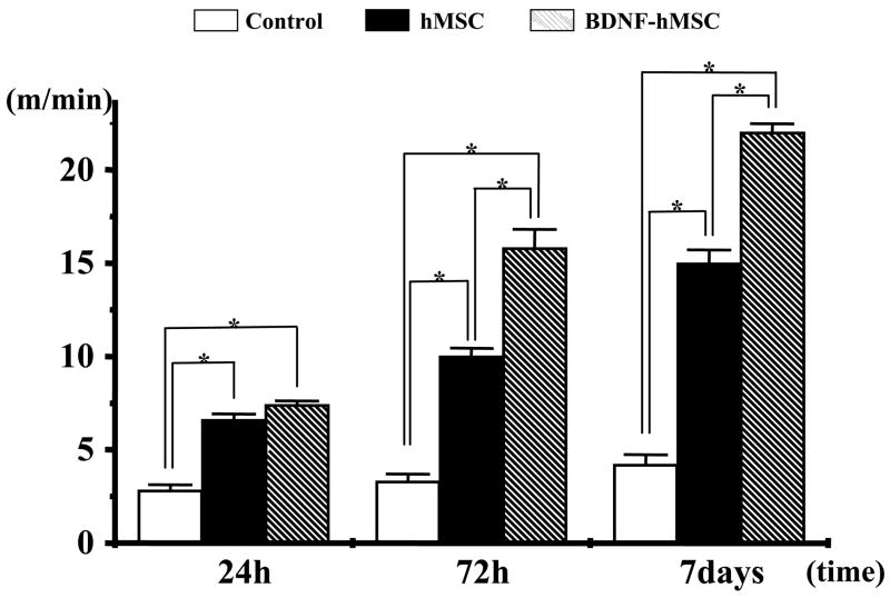
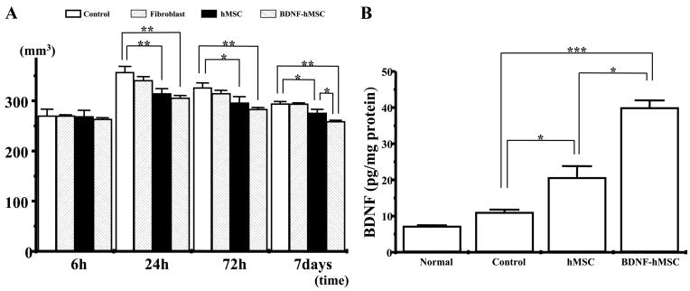
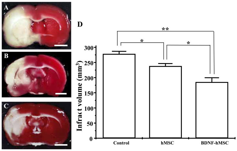
I.V. delivery of mesenchymal stem cells prepared from adult bone marrow reduces infarction size and ameliorates functional deficits in rat cerebral ischemia models. Administration of the brain-derived neurotrophic factor to the infarction site has also been demonstrated to be neuroprotective. To test the hypothesis that brain-derived neurotrophic factor contributes to the therapeutic benefits of mesenchymal stem cell delivery, we compared the efficacy of systemic delivery of human mesenchymal stem cells and human mesenchymal stem cells transfected with a fiber-mutant F/RGD adenovirus vector with a brain-derived neurotrophic factor gene (brain-derived neurotrophic factor-human mesenchymal stem cells). A permanent middle cerebral artery occlusion was induced by intraluminal vascular occlusion with a microfilament. Human mesenchymal stem cells and brain-derived neurotrophic factor-human mesenchymal stem cells were i.v. injected into the rats 6 h after middle cerebral artery occlusion. Lesion size was assessed at 6 h, 1, 3 and 7 days using MR imaging, and histological methods. Functional outcome was assessed using the treadmill stress test. Both human mesenchymal stem cells and brain-derived neurotrophic factor-human mesenchymal stem cells reduced lesion volume and elicited functional improvement compared with the control sham group, but the effect was greater in the brain-derived neurotrophic factor-human mesenchymal stem cell group. ELISA analysis of the infarcted hemisphere revealed an increase in brain-derived neurotrophic factor in the human mesenchymal stem cell groups, but a greater increase in the brain-derived neurotrophic factor-human mesenchymal stem cell group. These data support the hypothesis that brain-derived neurotrophic factor contributes to neuroprotection in cerebral ischemia and cellular delivery of brain-derived neurotrophic factor can be achieved by i.v. delivery of human mesenchymal stem cells.
静脉注射成年骨髓来源的间充质干细胞可减小大鼠脑缺血模型中的梗死面积并改善功能缺陷。向梗死部位给予脑源性神经营养因子也已被证明具有神经保护作用。为了验证脑源性神经营养因子有助于间充质干细胞递送的治疗益处这一假设,我们比较了人骨髓间充质干细胞和用纤维突变型F/RGD腺病毒载体转染了脑源性神经营养因子基因的人骨髓间充质干细胞(脑源性神经营养因子-人骨髓间充质干细胞)全身递送的效果。通过用微丝进行腔内血管闭塞诱导永久性大脑中动脉闭塞。在大脑中动脉闭塞6小时后,将人骨髓间充质干细胞和脑源性神经营养因子-人骨髓间充质干细胞静脉注射到大鼠体内。在6小时、1天、3天和7天时使用磁共振成像和组织学方法评估损伤大小。使用跑步机应激试验评估功能结局。与假手术对照组相比,人骨髓间充质干细胞和脑源性神经营养因子-人骨髓间充质干细胞均减小了损伤体积并引起功能改善,但在脑源性神经营养因子-人骨髓间充质干细胞组中效果更明显。对梗死半球的ELISA分析显示,人骨髓间充质干细胞组中脑源性神经营养因子增加,但在脑源性神经营养因子-人骨髓间充质干细胞组中增加更多。这些数据支持了脑源性神经营养因子有助于脑缺血神经保护的假设,并且通过静脉注射人骨髓间充质干细胞可以实现脑源性神经营养因子的细胞递送。