Szabo Zsolt, Xia Jinsong, Mathews William B, Brown Phillip R
Division of Nuclear Medicine, Johns Hopkins University School of Medicine, Baltimore, MD 21287, USA.
Semin Nucl Med. 2006 Jan;36(1):36-50. doi: 10.1053/j.semnuclmed.2005.08.003.
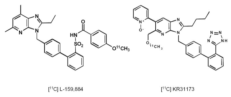
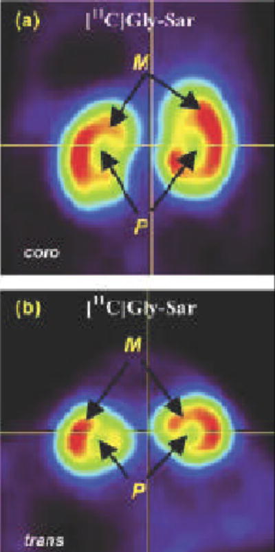
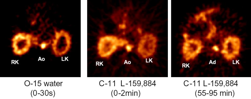
Positron emission tomography (PET) is perfectly suited for quantitative imaging of the kidneys, and the recent improvements in detector technology, computer hardware, and image processing software add to its appeal. Multiple positron emitting radioisotopes can be used for renal imaging. Some, including carbon-11, nitrogen-13, and oxygen-15, can be used at institutions with an on-site cyclotron. Other radioisotopes that may be even more useful in a clinical setting are those that either can be obtained from radionuclide generators (rubidium-82, copper-62) or have a sufficiently long half-life for transportation (fluorine-18). The clinical use of functional renal PET studies (blood flow, glomerular filtration rate) has been slow, in part because of the success of concurrent technologies, including single-photon emission computed tomography (SPECT) and planar gamma camera imaging. Renal blood flow studies can be performed with O-15-labeled water, N-13-labeled ammonia, rubidium-82, and copper-labeled PTSM. With these tracers, renal blood flow can be quantified using a modified microsphere kinetic model. Glomerular filtration can be imaged and quantified with gallium-68 EDTA or cobalt-55 EDTA. Measurements of renal blood flow with PET have potential applications in renovascular disease, in transplant rejection or acute tubular necrosis, in drug-induced nephropathies, ureteral obstruction, before and after revascularization, and before and after the placement of ureteral stents. The most important clinical application for imaging glomerular function with PET would be renovascular hypertension. Molecular imaging of the kidneys with PET is rather limited. At present, research is focused on the investigation of metabolism (acetate), membrane transporters (organic cation and anion transporters, pepT1 and pepT2, GLUT, SGLT), enzymes (ACE), and receptors (AT1R). Because many nephrological and urological disorders are initiated at the molecular and organelle levels and may remain localized at their origin for an extended period of time, new disease-specific molecular probes for PET studies of the kidneys need to be developed. Future applications of molecular renal imaging are likely to involve studies of tissue hypoxia and apoptosis in renovascular renal disease, renal cancer, and obstructive nephropathy, monitoring the molecular signatures of atherosclerotic plaques, measuring endothelial dysfunction and response to balloon revascularization and restenosis, molecular assessment of the nephrotoxic effects of cyclosporine, anticancer drugs, and radiation therapy. New radioligands will enhance the staging and follow-up of renal and prostate cancer. Methods will be developed for investigation of the kinetics of drug-delivery systems and delivery and deposition of prodrugs, reporter gene technology, delivery of gene therapy (nuclear and mitochondrial), assessment of the delivery of cellular, viral, and nonviral vectors (liposomes, polycations, fusion proteins, electroporation, hematopoietic stems cells). Of particular importance will be investigations of stem cell kinetics, including local presence, bloodborne migration, activation, seeding, and its role in renal remodeling (psychological, pathological, and therapy induced). Methods also could be established for investigating the role of receptors and oncoproteins in cellular proliferation, apoptosis, tubular atrophy, and interstitial fibrosis; monitoring ras gene targeting in kidney diseases, assessing cell therapy devices (bioartificial filters, renal tubule assist devices, and bioarticial kidneys), and targeting of signal transduction moleculas with growth factors and cytokines. These potential new approaches are, at best, in an experimental stage, and more research will be needed for their implementation.
正电子发射断层扫描(PET)非常适合肾脏的定量成像,并且探测器技术、计算机硬件和图像处理软件最近的改进增加了它的吸引力。多种发射正电子的放射性同位素可用于肾脏成像。其中一些,包括碳 - 11、氮 - 13和氧 - 15,可在配备现场回旋加速器的机构中使用。在临床环境中可能更有用的其他放射性同位素是那些可以从放射性核素发生器获得的(铷 - 82、铜 - 62)或具有足够长半衰期以便运输的(氟 - 18)。功能性肾脏PET研究(血流、肾小球滤过率)的临床应用进展缓慢,部分原因是同期技术的成功,包括单光子发射计算机断层扫描(SPECT)和平面伽马相机成像。肾脏血流研究可以用O - 15标记的水、N - 13标记的氨、铷 - 82和铜标记的PTSM进行。使用这些示踪剂,可以使用改良的微球动力学模型对肾脏血流进行定量。肾小球滤过可以用镓 - 68 EDTA或钴 - 55 EDTA进行成像和定量。PET测量肾脏血流在肾血管疾病、移植排斥或急性肾小管坏死、药物性肾病、输尿管梗阻、血管重建前后以及输尿管支架置入前后有潜在应用。PET成像肾小球功能最重要的临床应用是肾血管性高血压。PET对肾脏的分子成像相当有限。目前,研究集中在代谢(乙酸盐)、膜转运蛋白(有机阳离子和阴离子转运蛋白、肽转运体1和肽转运体2、葡萄糖转运蛋白、钠 - 葡萄糖协同转运蛋白)、酶(血管紧张素转换酶)和受体(血管紧张素Ⅱ1型受体)的研究上。因为许多肾病和泌尿系统疾病在分子和细胞器水平上起始,并且可能在其起源部位长时间局限存在,所以需要开发用于肾脏PET研究的新的疾病特异性分子探针。分子肾脏成像的未来应用可能涉及肾血管性肾病、肾癌和梗阻性肾病中组织缺氧和细胞凋亡的研究,监测动脉粥样硬化斑块的分子特征,测量内皮功能障碍以及对球囊血管重建和再狭窄的反应,环孢素、抗癌药物和放射治疗肾毒性作用的分子评估。新的放射性配体将提高肾癌和前列腺癌的分期及随访。将开发用于研究药物递送系统动力学以及前药递送和沉积、报告基因技术、基因治疗(核基因和线粒体基因)递送、评估细胞、病毒和非病毒载体(脂质体、聚阳离子、融合蛋白、电穿孔、造血干细胞)递送的方法。特别重要的将是干细胞动力学的研究,包括局部存在、血行迁移、激活、播种及其在肾脏重塑(生理、病理和治疗诱导)中的作用。还可以建立用于研究受体和癌蛋白在细胞增殖、凋亡、肾小管萎缩和间质纤维化中的作用的方法;监测肾脏疾病中ras基因靶向,评估细胞治疗装置(生物人工滤器、肾小管辅助装置和生物人工肾),以及用生长因子和细胞因子靶向信号转导分子。这些潜在的新方法充其量处于实验阶段,其实施还需要更多研究。