Olsen M L, Higashimori H, Campbell S L, Hablitz J J, Sontheimer H
Department of Neurobiology and Civitan International Research Center, University of Alabama at Birmingham, Birmingham, Alabama 35294, USA.
Glia. 2006 Apr 1;53(5):516-28. doi: 10.1002/glia.20312.
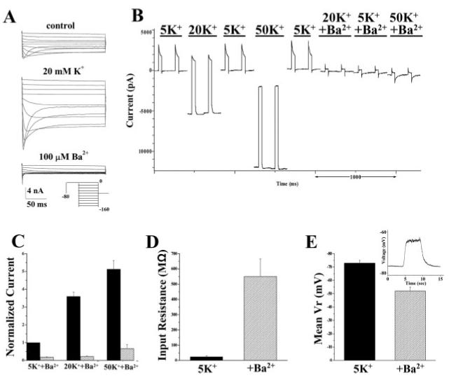
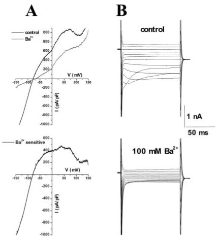
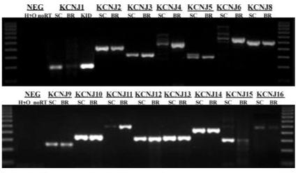
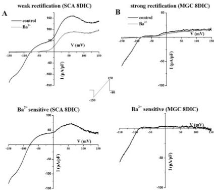
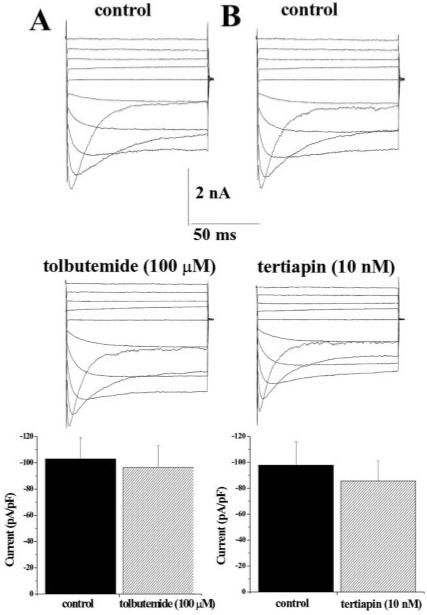
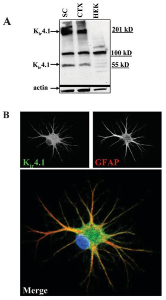
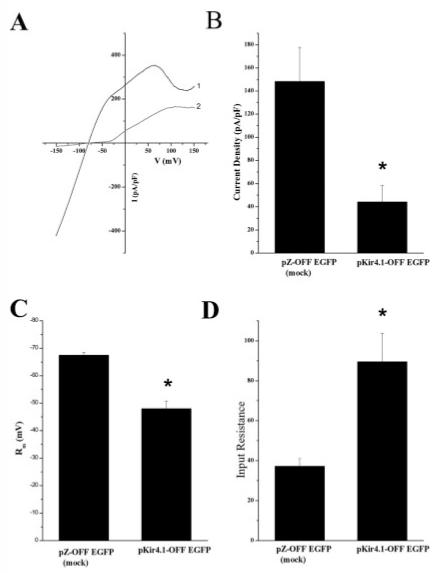
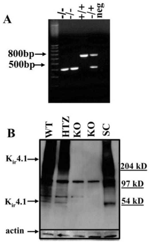
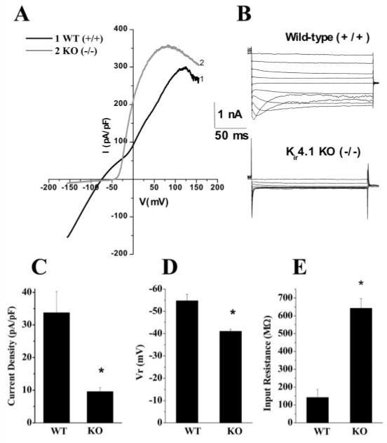
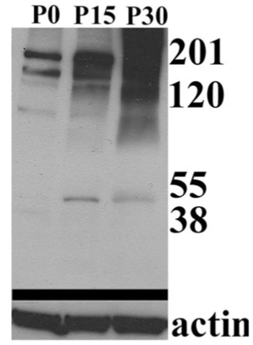
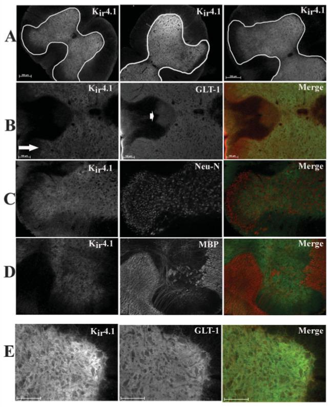
Spinal cord astrocytes (SCA) have a high permeability to K+ and hence have hyperpolarized resting membrane potentials. The underlying K+ channels are believed to participate in the uptake of neuronally released K+. These K+ channels have been studied extensively with regard to their biophysics and pharmacology, but their molecular identity in spinal cord is currently unknown. Using a combination of approaches, we demonstrate that channels composed of the Kir4.1 subunit are responsible for mediating the resting K+ conductance in SCA. Biophysical analysis demonstrates astrocytic Kir currents as weakly rectifying, potentiated by increasing [K+]o, and inhibited by micromolar concentrations of Ba2+. These currents were insensitive to tolbutemide, a selective blocker of Kir6.x channels, and to tertiapin, a blocker for Kir1.1 and Kir3.1/3.4 channels. PCR and Western blot analysis show prominent expression of Kir4.1 in SCA, and immunocytochemistry shows localization Kir4.1 channels to the plasma membrane. Kir4.1 protein levels show a developmental upregulation in vivo that parallels an increase in currents recorded over the same time period. Kir4.1 is highly expressed throughout most areas of the gray matter in spinal cord in vivo and recordings from spinal cord slices show prominent Kir currents. Electrophysiological recordings comparing SCA of wild-type mice with those of homozygote Kir4.1 knockout mice confirm a complete and selective absence of Kir channels in the knockout mice, suggesting that Kir4.1 is the principle channel mediating the resting K+ conductance in SCA in vitro and in situ.
脊髓星形胶质细胞(SCA)对K⁺具有高通透性,因此具有超极化的静息膜电位。据信,其潜在的K⁺通道参与摄取神经元释放的K⁺。这些K⁺通道在生物物理学和药理学方面已得到广泛研究,但其在脊髓中的分子身份目前尚不清楚。通过多种方法相结合,我们证明由Kir4.1亚基组成的通道负责介导SCA中的静息K⁺电导。生物物理分析表明,星形胶质细胞的Kir电流呈弱整流性,随着细胞外K⁺浓度([K⁺]o)升高而增强,并受到微摩尔浓度Ba²⁺的抑制。这些电流对Kir6.x通道的选择性阻滞剂甲苯磺丁脲以及Kir1.1和Kir3.1/3.4通道的阻滞剂特律平不敏感。PCR和蛋白质印迹分析显示Kir4.1在SCA中显著表达,免疫细胞化学显示Kir4.1通道定位于质膜。Kir4.1蛋白水平在体内呈现发育性上调,这与同一时期记录的电流增加情况平行。在体内,Kir4.1在脊髓灰质的大部分区域中高度表达,并且从脊髓切片记录显示出明显的Kir电流。将野生型小鼠的SCA与纯合子Kir4.1基因敲除小鼠的SCA进行电生理记录比较,证实基因敲除小鼠中完全且选择性地缺失Kir通道,这表明Kir4.1是在体外和原位介导SCA中静息K⁺电导的主要通道。