• 文献检索
  • 文档翻译
  • 深度研究
  • 学术资讯
  • Suppr Zotero 插件Zotero 插件
  • 邀请有礼
  • 套餐&价格
  • 历史记录
应用&插件
Suppr Zotero 插件Zotero 插件浏览器插件Mac 客户端Windows 客户端微信小程序
定价
高级版会员购买积分包购买API积分包
服务
文献检索文档翻译深度研究API 文档MCP 服务
关于我们
关于 Suppr公司介绍联系我们用户协议隐私条款
关注我们

Suppr 超能文献

核心技术专利:CN118964589B侵权必究
粤ICP备2023148730 号-1Suppr @ 2026

文献检索

告别复杂PubMed语法,用中文像聊天一样搜索,搜遍4000万医学文献。AI智能推荐,让科研检索更轻松。

立即免费搜索

文件翻译

保留排版,准确专业,支持PDF/Word/PPT等文件格式,支持 12+语言互译。

免费翻译文档

深度研究

AI帮你快速写综述,25分钟生成高质量综述,智能提取关键信息,辅助科研写作。

立即免费体验

An electron microscopic study of virus particles in rhesus monkey placenta.

作者信息

Feldman D

出版信息

Proc Natl Acad Sci U S A. 1975 Jan;72(1):118-21. doi: 10.1073/pnas.72.1.118.

DOI:10.1073/pnas.72.1.118
PMID:164011
原文链接:https://pmc.ncbi.nlm.nih.gov/articles/PMC432252/
Abstract

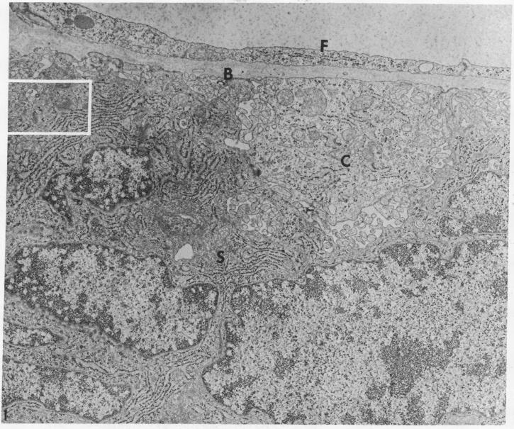
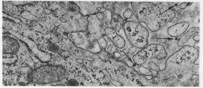
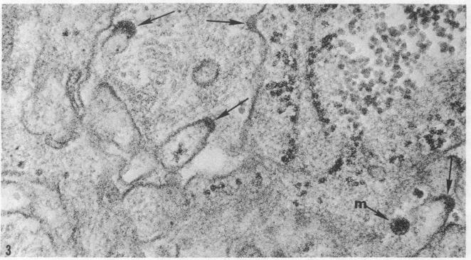
Examination of chorionic villi from rhesus monkey placenta revealed the presence of C-type virus particles budding from syncytial trophoblast, pericytes, Hofbauer cells, and mesenchyme. In addition, particles, were found budding from cells of the cytotrophoblastic cell column and decidual basalis. They measured 30 nm in diameter, had a dense central core, surrounded by a narrow, electron-lucent zone, and were enclosed by an outer unit membrane.

摘要
https://cdn.ncbi.nlm.nih.gov/pmc/blobs/c947/432252/c5b28e4ab711/pnas00044-0126-c.jpg
https://cdn.ncbi.nlm.nih.gov/pmc/blobs/c947/432252/ec4c639466b1/pnas00044-0125-a.jpg
https://cdn.ncbi.nlm.nih.gov/pmc/blobs/c947/432252/89eefabfdee5/pnas00044-0125-b.jpg
https://cdn.ncbi.nlm.nih.gov/pmc/blobs/c947/432252/acd036daba10/pnas00044-0126-a.jpg
https://cdn.ncbi.nlm.nih.gov/pmc/blobs/c947/432252/41b942dc6111/pnas00044-0126-b.jpg
https://cdn.ncbi.nlm.nih.gov/pmc/blobs/c947/432252/c5b28e4ab711/pnas00044-0126-c.jpg

相似文献

1
An electron microscopic study of virus particles in rhesus monkey placenta.
Proc Natl Acad Sci U S A. 1975 Jan;72(1):118-21. doi: 10.1073/pnas.72.1.118.
2
Virus particles in the basal plate of rhesus monkey and baboon placenta.恒河猴和狒狒胎盘基底板中的病毒颗粒。
Cancer Res. 1979 May;39(5):1772-83.
3
Developmental changes in the fine structure of the chorion laeve (smooth chorion) of the rhesus monkey placenta.恒河猴胎盘平滑绒毛膜精细结构的发育变化
Anat Rec. 1981 Jun;200(2):163-75. doi: 10.1002/ar.1092000206.
4
C-type virus particles in placentas of rhesus monkeys after maternal treatment with recombinant leukocyte A interferon.
J Exp Pathol. 1989 Fall;4(4):193-8.
5
Preferential expression of endogenous type C viral antigen in Rhesus placenta during ontogenesis.恒河猴胎盘在个体发育过程中内源性C型病毒抗原的优先表达。
Virology. 1981 Jul 15;112(1):365-9. doi: 10.1016/0042-6822(81)90644-9.
6
Efficient isolation of endogenous rhesus retrovirus from trophoblast.从滋养层细胞中高效分离内源性恒河猴逆转录病毒。
Virology. 1983 Jul 30;128(2):518-23. doi: 10.1016/0042-6822(83)90281-7.
7
Ultrastructure of the placental villi, chorion laeve, and decidua parietalis in normal and hypertensive pregnant women.正常孕妇和高血压孕妇胎盘绒毛、平滑绒毛膜及子宫壁蜕膜的超微结构
Am J Obstet Gynecol. 1977 Nov 1;129(5):492-8. doi: 10.1016/0002-9378(77)90085-0.
8
Human chorionic trophoblasts, decidual cells, and macrophages: a histochemical and electron microscopic study.
Am J Obstet Gynecol. 1981 Jun 1;140(3):261-8. doi: 10.1016/0002-9378(81)90271-4.
9
The chorioallantoic placenta of the spotted hyena (Crocuta crocuta Erxleben): an electron microscopic study.斑鬣狗(克罗克斑鬣狗,学名:Crocuta crocuta Erxleben)的绒膜尿囊胎盘:一项电子显微镜研究
Anat Rec. 1982 Nov;204(3):215-22. doi: 10.1002/ar.1092040306.
10
Developmental changes in rhesus monkey placental villi and cell columns. Scanning electron microscopy.恒河猴胎盘绒毛和细胞柱的发育变化。扫描电子显微镜观察。
Anat Embryol (Berl). 1982 Dec;165(3):361-76. doi: 10.1007/BF00305573.

引用本文的文献

1
Extrachromosomal elements as possible agents of adaptation and development.作为适应与发育潜在因子的染色体外元件
Bacteriol Rev. 1976 Sep;40(3):552-90. doi: 10.1128/br.40.3.552-590.1976.
2
C-type virus particle formation in erythroblastic islands in spleens of C3Hf mice injected with erythropoietin.注射促红细胞生成素的C3Hf小鼠脾脏中红细胞生成岛的C型病毒颗粒形成。
Proc Natl Acad Sci U S A. 1976 Oct;73(10):3710-3. doi: 10.1073/pnas.73.10.3710.
3
The occurrence and frequency of type C virus-like particles in placentas from patients with systemic lupus erythematosus and from normal subjects.

本文引用的文献

1
Brief communication: C-type particles in normal human placentas.简短通讯:正常人胎盘组织中的C型颗粒
J Natl Cancer Inst. 1973 Apr;50(4):1081-4. doi: 10.1093/jnci/50.4.1081.
2
Observations of apparent C-type particles in baboon (Papio cynocephalus) placentas.在狒狒(Papio cynocephalus)胎盘内观察到明显的C型颗粒。
Science. 1973 Mar 30;179(4080):1332-3. doi: 10.1126/science.179.4080.1332.
3
Ultrastructural comparison of placental virus with several type-c- oncogenic viruses.胎盘病毒与几种C型致癌病毒的超微结构比较。
系统性红斑狼疮患者及正常受试者胎盘组织中C型病毒样颗粒的出现情况及频率
Am J Pathol. 1976 May;83(2):383-94.
4
C-type virus particles in placenta of normal healthy Sprague-Dawley rats.正常健康的斯普拉格-道利大鼠胎盘内的C型病毒颗粒。
Proc Natl Acad Sci U S A. 1975 Aug;72(8):3240-4. doi: 10.1073/pnas.72.8.3240.
5
Intracisternal virus-like particles in a human pituitary adenoma.
Z Krebsforsch Klin Onkol Cancer Res Clin Oncol. 1976 Feb 25;85(2):111-5. doi: 10.1007/BF00304943.
J Natl Cancer Inst. 1974 Apr;52(4):1379-81. doi: 10.1093/jnci/52.4.1379.
4
C-type virus particles in placentas and fetal tissues of Rhesus monkeys.恒河猴胎盘和胎儿组织中的C型病毒颗粒。
J Natl Cancer Inst. 1973 Jul;51(1):225-33. doi: 10.1093/jnci/51.1.225.
5
Electron microscopic study of the mouse leukemia virus (Gross) in organs of mouse embryos from virus-injected and normal C3Hf parents.对来自注射病毒的和正常C3Hf亲代的小鼠胚胎器官中(格罗斯)小鼠白血病病毒的电子显微镜研究。
Cancer Res. 1967 Oct;27(10):1792-804.