• 文献检索
  • 文档翻译
  • 深度研究
  • 学术资讯
  • Suppr Zotero 插件Zotero 插件
  • 邀请有礼
  • 套餐&价格
  • 历史记录
应用&插件
Suppr Zotero 插件Zotero 插件浏览器插件Mac 客户端Windows 客户端微信小程序
定价
高级版会员购买积分包购买API积分包
服务
文献检索文档翻译深度研究API 文档MCP 服务
关于我们
关于 Suppr公司介绍联系我们用户协议隐私条款
关注我们

Suppr 超能文献

核心技术专利:CN118964589B侵权必究
粤ICP备2023148730 号-1Suppr @ 2026

文献检索

告别复杂PubMed语法,用中文像聊天一样搜索,搜遍4000万医学文献。AI智能推荐,让科研检索更轻松。

立即免费搜索

文件翻译

保留排版,准确专业,支持PDF/Word/PPT等文件格式,支持 12+语言互译。

免费翻译文档

深度研究

AI帮你快速写综述,25分钟生成高质量综述,智能提取关键信息,辅助科研写作。

立即免费体验

牛副流感3型病毒感染:病毒在牛胚胎细胞培养物中的复制及通过速率区带离心法分离病毒粒子

Bovine parainfluenza type 3 virus infection: virus replication in bovine embryonic cell cultures and virion separation by rate-zonal centrifugation.

作者信息

Tsai K S, Thomson R G

出版信息

Infect Immun. 1975 Apr;11(4):770-82. doi: 10.1128/iai.11.4.770-782.1975.

DOI:10.1128/iai.11.4.770-782.1975
PMID:164407
原文链接:https://pmc.ncbi.nlm.nih.gov/articles/PMC415136/
Abstract

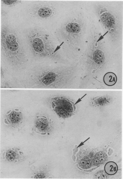
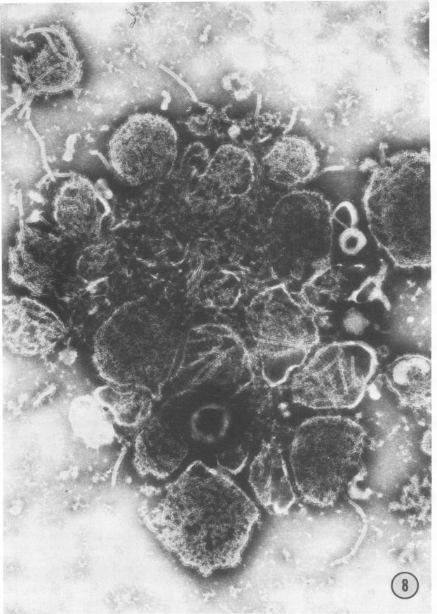
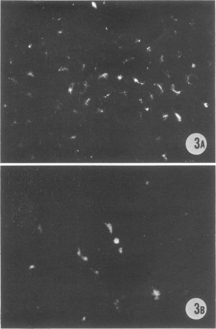
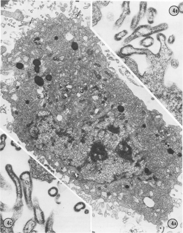
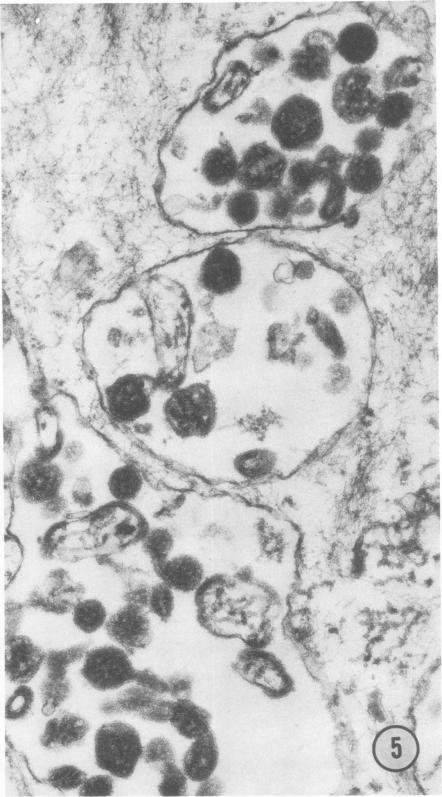
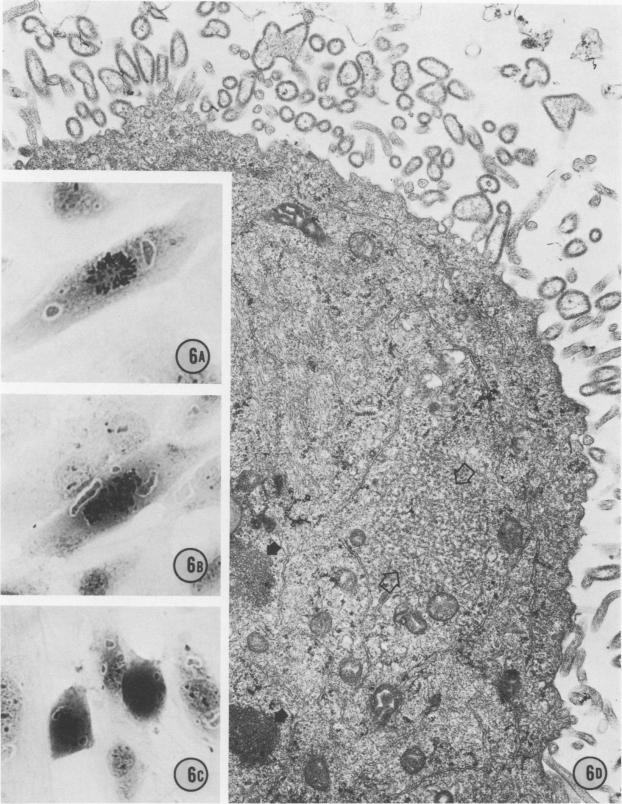
Replicative sequences of a bovine strain of parainfluenza type 3 virus in bovine embryonic kidney and spleen cell cultures were investigated by light and fluorescence microscopy and by ultrathin section and negative-contrast electron microscopy. Observations from light and fluorescence microscopy showed that intracytoplasmic inclusions were detected as small granules surrounding the nuclei of more than 90 percent of the cell population by day 2 postinoculation. With the increase of postexposure times, these inclusions coalesced into larger bodies which occupied large portions of the cell. Ultrastructurally, the first sign of virus development was the appearance of aggregates of viral nucleocapsids in the vicinity of the nucleus. With the concomitant accumulation of viral nucleocapsids in the cytoplasm, the virus maturation was expressed by budding processes through the cell membrane into round, oval, or elongated forms. Eosinophilic inclusions were demonstrable in many mitotic cells. Ultrastructurally, these cells were observed to produce virus particles by a process identical to that of resting cells. Virions, prepared from infected culture fluid and negatively stained, appeared to be pleomorphic and their diameter ranged from 200 to 600 mm. The virions were separated, by rate-zonal centrifugation, into two subclasses in a sucrose gradient (15 to 60 percent, wt/wt). The slowly sedimenting virions had a density approximately 1.20 gm/cm3 and an average size of 200 nm in diameter, whereas the faster-sedimenting virions had a density of 1.24 gm/cm3 and average diameter of 400 nm.

摘要

利用光学显微镜、荧光显微镜、超薄切片及负染色电子显微镜对牛副流感3型病毒牛株在牛胚胎肾和脾细胞培养物中的复制序列进行了研究。光学显微镜和荧光显微镜观察结果显示,接种后第2天,超过90%的细胞群体中,细胞核周围的胞质内包涵体被检测为小颗粒。随着暴露后时间的增加,这些包涵体融合成更大的体块,占据了细胞的大部分区域。在超微结构上,病毒发育的第一个迹象是细胞核附近出现病毒核衣壳聚集体。随着病毒核衣壳在细胞质中的积累,病毒成熟表现为通过细胞膜出芽形成圆形、椭圆形或细长形。在许多有丝分裂细胞中可显示嗜酸性包涵体。在超微结构上,观察到这些细胞通过与静止细胞相同的过程产生病毒颗粒。从感染的培养液中制备并经负染色的病毒粒子似乎呈多形性,其直径范围为200至600毫米。通过速率区带离心,病毒粒子在蔗糖梯度(15%至60%,重量/重量)中被分为两个亚类。沉降较慢的病毒粒子密度约为1.20克/立方厘米,平均直径为200纳米,而沉降较快的病毒粒子密度为1.24克/立方厘米,平均直径为400纳米。

https://cdn.ncbi.nlm.nih.gov/pmc/blobs/036a/415136/660a87aa57a4/iai00232-0172-a.jpg
https://cdn.ncbi.nlm.nih.gov/pmc/blobs/036a/415136/fc892d567bfa/iai00232-0165-a.jpg
https://cdn.ncbi.nlm.nih.gov/pmc/blobs/036a/415136/1bca2ded34de/iai00232-0166-a.jpg
https://cdn.ncbi.nlm.nih.gov/pmc/blobs/036a/415136/1afe5694eabc/iai00232-0167-a.jpg
https://cdn.ncbi.nlm.nih.gov/pmc/blobs/036a/415136/dc8eabeffce1/iai00232-0168-a.jpg
https://cdn.ncbi.nlm.nih.gov/pmc/blobs/036a/415136/55da1129b66f/iai00232-0169-a.jpg
https://cdn.ncbi.nlm.nih.gov/pmc/blobs/036a/415136/a9131fdf7629/iai00232-0170-a.jpg
https://cdn.ncbi.nlm.nih.gov/pmc/blobs/036a/415136/660a87aa57a4/iai00232-0172-a.jpg

相似文献

1
Bovine parainfluenza type 3 virus infection: virus replication in bovine embryonic cell cultures and virion separation by rate-zonal centrifugation.牛副流感3型病毒感染:病毒在牛胚胎细胞培养物中的复制及通过速率区带离心法分离病毒粒子
Infect Immun. 1975 Apr;11(4):770-82. doi: 10.1128/iai.11.4.770-782.1975.
2
Bovine parainfluenza type 3 virus infection: ultrastructural aspects of viral pathogenesis in the bovine respiratory tract.牛副流感3型病毒感染:牛呼吸道病毒发病机制的超微结构方面
Infect Immun. 1975 Apr;11(4):783-803. doi: 10.1128/iai.11.4.783-803.1975.
3
Mumps virus replication in chick embryo lung cells: properties of ribonucleic acids in virions and infected cells.腮腺炎病毒在鸡胚肺细胞中的复制:病毒粒子及受感染细胞中核糖核酸的特性
J Virol. 1971 Aug;8(2):161-73. doi: 10.1128/JVI.8.2.161-173.1971.
4
Characterization of a calf diarrheal coronavirus.一种犊牛腹泻冠状病毒的特性分析
Am J Vet Res. 1976 Sep;37(9):1031-41.
5
Pathogenesis of bovine parainfluenza 3 virus infection in organ cultures.牛副流感3型病毒在器官培养物中的致病机制。
J Comp Pathol. 1969 Jul;79(3):347-54. doi: 10.1016/0021-9975(69)90049-8.
6
Isolation and partial characterization of a novel paramyxovirus from the gills of diseased seawater-reared Atlantic salmon (Salmo salar L).从患病海水养殖大西洋鲑(Salmo salar L)鳃中分离出一种新型副粘病毒并进行部分特性分析。
J Gen Virol. 2003 Aug;84(Pt 8):2179-2189. doi: 10.1099/vir.0.18962-0.
7
Further observations on the characteristics of a bovine parainfluenza-3 variant.关于一种牛副流感3型变体特征的进一步观察
Can J Comp Med. 1975 Jan;39(1):76-9.
8
Cytopathic effects and immunofluorescence produced by the J 121 strain of bovine Parainfluenza 3 virus in tissue culture.牛副流感3病毒J 121株在组织培养中产生的细胞病变效应和免疫荧光
J Comp Pathol. 1965 Jul;75(3):287-97. doi: 10.1016/0021-9975(65)90034-4.
9
Virological examination of bovine mammary lymph nodes.牛乳腺淋巴结的病毒学检查
Can J Comp Med. 1970 Oct;34(4):354-5.
10
Parainfluenza-5 virus. Infection of man and animal.
Prog Med Virol. 1972;14:241-74.

引用本文的文献

1
Analysis of bovine parainfluenza-3 virus replication in bovine embryonic lung cells by indirect fluorescent antibody and hemadsorption assays.通过间接荧光抗体和血细胞吸附试验分析牛副流感3型病毒在牛胚胎肺细胞中的复制情况。
J Virol Methods. 1990 Jan;27(1):113-9. doi: 10.1016/0166-0934(90)90151-5.
2
Characterization of bovine parainfluenza virus type 3.牛副流感3型病毒的特性分析
Microbiol Immunol. 1979;23(7):617-28. doi: 10.1111/j.1348-0421.1979.tb00502.x.
3
Replication of parainfluenza type 3 virus in alveolar macrophages: evidence of in vivo infection and of in vitro temperature sensitivity in virus maturation.

本文引用的文献

1
THE COMBINED EFFECT OF PASTEURELLA MULTOCIDA AND MYXOVIRUS PARAINFLUENZA-3 UPON CALVES.多杀性巴氏杆菌与副流感病毒3型对犊牛的联合作用
Am J Vet Res. 1963 Sep;24:939-47.
2
The fine structure of the parainfluenza viruses.副流感病毒的精细结构。
Arch Gesamte Virusforsch. 1962;12:138-42. doi: 10.1007/BF01258760.
3
A carrier state of mumps virus in human conjunctiva cells. I. General characteristics.人类结膜细胞中腮腺炎病毒的携带状态。I. 一般特征。
3型副流感病毒在肺泡巨噬细胞中的复制:体内感染及病毒成熟过程中体外温度敏感性的证据。
Infect Immun. 1977 Dec;18(3):780-91. doi: 10.1128/iai.18.3.780-791.1977.
4
Bovine parainfluenza type 3 virus infection: ultrastructural aspects of viral pathogenesis in the bovine respiratory tract.牛副流感3型病毒感染:牛呼吸道病毒发病机制的超微结构方面
Infect Immun. 1975 Apr;11(4):783-803. doi: 10.1128/iai.11.4.783-803.1975.
J Exp Med. 1962 Nov 1;116(5):739-50. doi: 10.1084/jem.116.5.739.
4
[Electron microscopy studies on the behavior of a parainfluenza-3 virus isolated from calves in calf kidney cell culture].[从犊牛分离的副流感3型病毒在犊牛肾细胞培养中的行为的电子显微镜研究]
Arch Gesamte Virusforsch. 1962;12:404-20.
5
Experimental transmission of shipping fever in calves.犊牛运输热的实验性传播
Am J Vet Res. 1963 Mar;24:287-94.
6
Studies on the transmission and etiology of bovine shipping fever.牛运输热的传播与病因学研究。
Am J Vet Res. 1962 May;23:548-53.
7
The development of Newcastle disease virus in cells of the chorio-allantoic membrane as studied by thin section.通过超薄切片研究新城疫病毒在尿囊绒毛膜细胞中的发育。
Bull Johns Hopkins Hosp. 1953 Apr;92(4):309-29.
8
Density separable fractions during growth of measles virus.麻疹病毒生长过程中的密度可分离组分。
J Immunol. 1966 Oct;97(4):458-69.
9
Ultrastructure of measles virus in cultures of hamster cerebellum.仓鼠小脑培养物中麻疹病毒的超微结构
J Virol. 1969 Aug;4(2):169-81. doi: 10.1128/JVI.4.2.169-181.1969.
10
Sendai virus replication: an ultrastructural comparison of productive and abortive infections in avian cells.仙台病毒复制:禽类细胞中增殖性感染与流产性感染的超微结构比较
J Gen Virol. 1970 Dec;9(3):169-77. doi: 10.1099/0022-1317-9-3-169.