Dancause Numa, Barbay Scott, Frost Shawn B, Plautz Erik J, Stowe Ann M, Friel Kathleen M, Nudo Randolph J
Department of Molecular and Integrative Physiology, University of Kansas Medical Center, Kansas City, Kansas 66160, USA.
J Comp Neurol. 2006 Apr 1;495(4):374-90. doi: 10.1002/cne.20875.
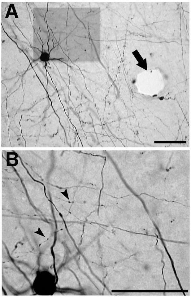
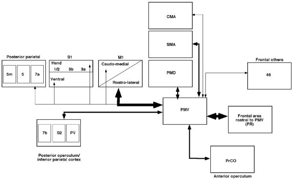
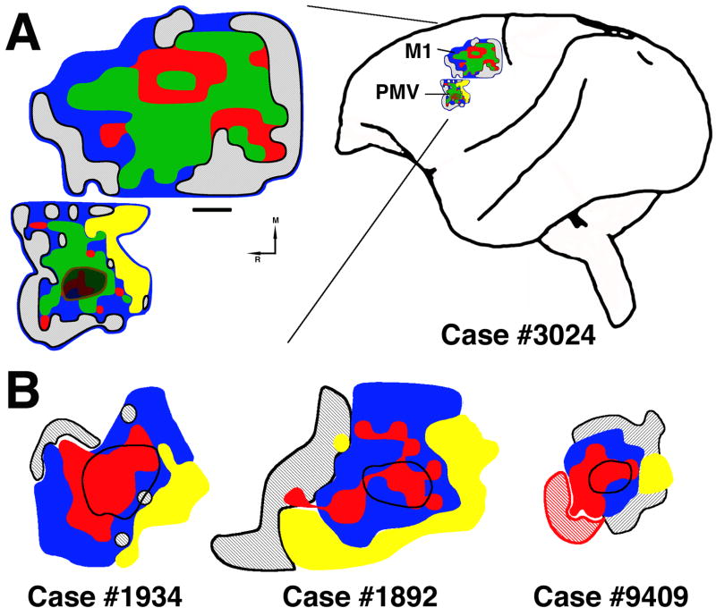
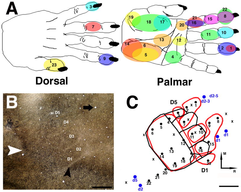
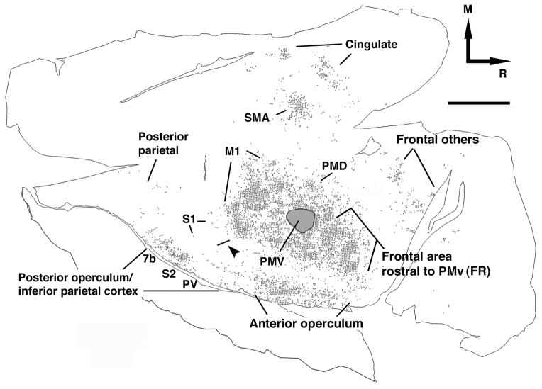
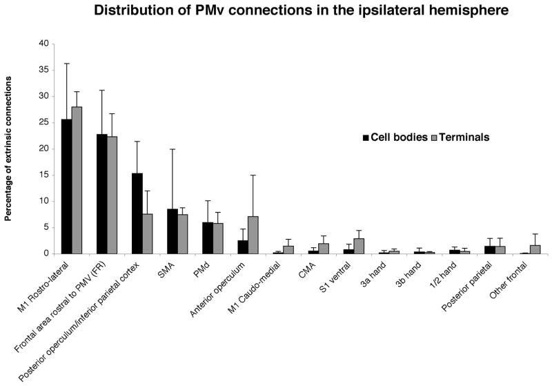
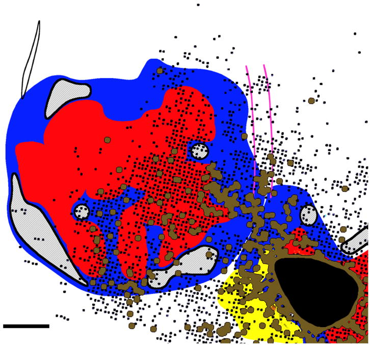
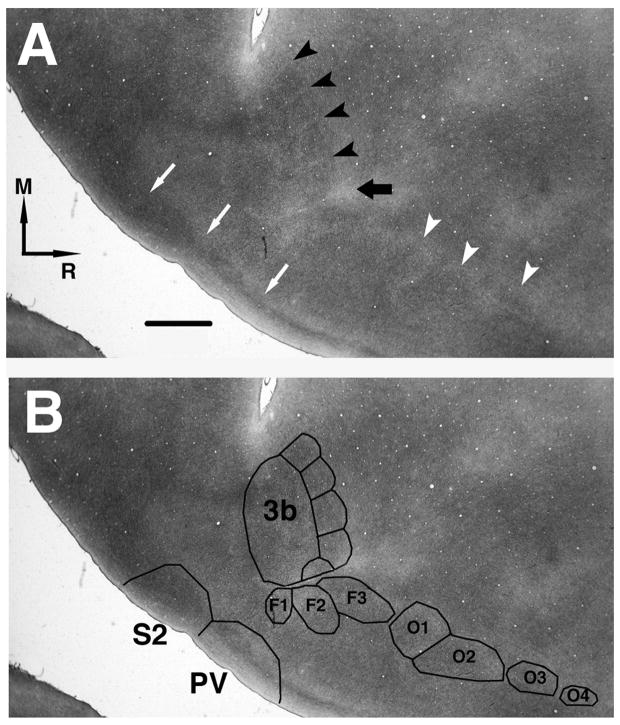
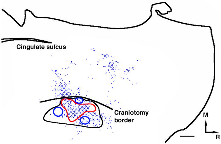
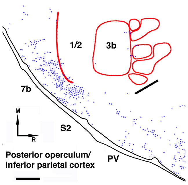
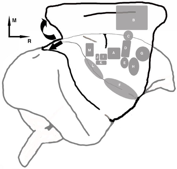
The present study describes the pattern of connections of the ventral premotor cortex (PMv) with various cortical regions of the ipsilateral hemisphere in adult squirrel monkeys. Particularly, we 1) quantified the proportion of inputs and outputs that the PMv distal forelimb representation shares with other areas in the ipsilateral cortex and 2) defined the pattern of PMv connections with respect to the location of the distal forelimb representation in primary motor cortex (M1), primary somatosensory cortex (S1), and supplementary motor area (SMA). Intracortical microstimulation techniques (ICMS) were used in four experimentally naïve monkeys to identify M1, PMv, and SMA forelimb movement representations. Multiunit recording techniques and myelin staining were used to identify the S1 hand representation. Then, biotinylated dextran amine (BDA; 10,000 MW) was injected in the center of the PMv distal forelimb representation. After tangential sectioning, the distribution of BDA-labeled cell bodies and terminal boutons was documented. In M1, labeling followed a rostrolateral pattern, largely leaving the caudomedial M1 unlabeled. Quantification of somata and terminals showed that two areas share major connections with PMv: M1 and frontal areas immediately rostral to PMv, designated as frontal rostral area (FR). Connections with this latter region have not been described previously. Moderate connections were found with PMd, SMA, anterior operculum, and posterior operculum/inferior parietal area. Minor connections were found with diverse areas of the precentral and parietal cortex, including S1. No statistical difference between the proportions of inputs and outputs for any location was observed, supporting the reciprocity of PMv intracortical connections.
本研究描述了成年松鼠猴腹侧运动前皮层(PMv)与同侧半球各皮层区域的连接模式。具体而言,我们1)量化了PMv远侧前肢代表区与同侧皮层其他区域共享的输入和输出比例,以及2)根据初级运动皮层(M1)、初级躯体感觉皮层(S1)和辅助运动区(SMA)中远侧前肢代表区的位置,确定了PMv的连接模式。对4只未经实验的猴子采用皮层内微刺激技术(ICMS)来识别M1、PMv和SMA的前肢运动代表区。采用多单位记录技术和髓鞘染色来识别S1的手部代表区。然后,将生物素化葡聚糖胺(BDA;分子量10,000)注入PMv远侧前肢代表区的中心。在进行切线切片后,记录BDA标记的细胞体和终末扣结的分布。在M1中,标记呈 rostrolateral 模式,大部分使M1的 caudomedial 区域未被标记。对细胞体和终末的量化显示,有两个区域与PMv有主要连接:M1和紧接在PMv前方的额叶区域,称为额前区(FR)。与后一区域的连接此前尚未有描述。发现与PMd、SMA、前岛盖和后岛盖/顶下区有中等程度的连接。与中央前回和顶叶皮层的不同区域,包括S1,有少量连接。未观察到任何位置的输入和输出比例之间存在统计学差异,这支持了PMv皮层内连接的相互性。