• 文献检索
  • 文档翻译
  • 深度研究
  • 学术资讯
  • Suppr Zotero 插件Zotero 插件
  • 邀请有礼
  • 套餐&价格
  • 历史记录
应用&插件
Suppr Zotero 插件Zotero 插件浏览器插件Mac 客户端Windows 客户端微信小程序
定价
高级版会员购买积分包购买API积分包
服务
文献检索文档翻译深度研究API 文档MCP 服务
关于我们
关于 Suppr公司介绍联系我们用户协议隐私条款
关注我们

Suppr 超能文献

核心技术专利:CN118964589B侵权必究
粤ICP备2023148730 号-1Suppr @ 2026

文献检索

告别复杂PubMed语法,用中文像聊天一样搜索,搜遍4000万医学文献。AI智能推荐,让科研检索更轻松。

立即免费搜索

文件翻译

保留排版,准确专业,支持PDF/Word/PPT等文件格式,支持 12+语言互译。

免费翻译文档

深度研究

AI帮你快速写综述,25分钟生成高质量综述,智能提取关键信息,辅助科研写作。

立即免费体验

相似文献

1
An instrumented scaffold can monitor loading in the knee joint.一种带有监测装置的支架可以监测膝关节的负荷情况。
J Biomed Mater Res B Appl Biomater. 2006 Nov;79(2):218-28. doi: 10.1002/jbm.b.30532.
2
Sensate scaffolds can reliably detect joint loading.感觉性支架能够可靠地检测关节负荷。
J Biomed Mater Res B Appl Biomater. 2007 Apr;81(1):30-9. doi: 10.1002/jbm.b.30632.
3
Sensate scaffolds coupled to telemetry can monitor in vivo loading from within a joint over extended periods of time.与遥测技术相结合的传感支架能够在较长时间内从关节内部监测体内负荷。
J Biomed Mater Res B Appl Biomater. 2008 Jan;84(1):263-70. doi: 10.1002/jbm.b.30869.
4
Successful Total Meniscus Reconstruction Using a Novel Fiber-Reinforced Scaffold: A 16- and 32-Week Study in an Ovine Model.使用新型纤维增强支架成功进行全半月板重建:在绵羊模型中的16周和32周研究。
Am J Sports Med. 2015 Oct;43(10):2528-37. doi: 10.1177/0363546515595065. Epub 2015 Aug 21.
5
Aptamer-Functionalized Bioscaffold Enhances Cartilage Repair by Improving Stem Cell Recruitment in Osteochondral Defects of Rabbit Knees.适配体功能化生物支架通过改善兔膝关节骨软骨缺损中的干细胞募集来增强软骨修复。
Am J Sports Med. 2019 Aug;47(10):2316-2326. doi: 10.1177/0363546519856355. Epub 2019 Jun 24.
6
In vivo evaluation of 3-dimensional polycaprolactone scaffolds for cartilage repair in rabbits.兔体内评估用于软骨修复的 3 维聚己内酯支架。
Am J Sports Med. 2010 Mar;38(3):509-19. doi: 10.1177/0363546509352448. Epub 2010 Jan 21.
7
Time evolution of in vivo articular cartilage repair induced by bone marrow stimulation and scaffold implantation in rabbits.兔骨髓刺激和支架植入诱导的体内关节软骨修复的时间演变
Int J Artif Organs. 2015 Apr;38(4):210-23. doi: 10.5301/ijao.5000404. Epub 2015 Apr 29.
8
Autologous-cell-derived, tissue-engineered cartilage for repairing articular cartilage lesions in the knee: study protocol for a randomized controlled trial.用于修复膝关节软骨损伤的自体细胞来源组织工程软骨:一项随机对照试验的研究方案
Trials. 2017 Nov 6;18(1):519. doi: 10.1186/s13063-017-2251-6.
9
Load measurement accuracy from sensate scaffolds with and without a cartilage surface.
J Invest Surg. 2010 Jun;23(3):156-62. doi: 10.3109/08941939.2010.481006.
10
[Arthroscopic tissue engineering scaffold repair for cartilage injuries].[关节镜下组织工程支架修复软骨损伤]
Beijing Da Xue Xue Bao Yi Xue Ban. 2025 Apr 18;57(2):384-387. doi: 10.19723/j.issn.1671-167X.2025.02.025.

引用本文的文献

1
Implantable sensor technology: measuring bone and joint biomechanics of daily life in vivo.可植入传感器技术:测量日常生活中体内骨骼和关节的生物力学。
Arthritis Res Ther. 2013 Jan 31;15(1):203. doi: 10.1186/ar4138.
2
Knee joint forces: prediction, measurement, and significance.膝关节力:预测、测量及意义
Proc Inst Mech Eng H. 2012 Feb;226(2):95-102. doi: 10.1177/0954411911433372.
3
A handheld computer as part of a portable in vivo knee joint load monitoring system.作为便携式体内膝关节负荷监测系统一部分的手持计算机。
J Med Device. 2008 Sep 1;2(3):350011-350019. doi: 10.1115/1.2952815.
4
A novel biomimetic polymer scaffold design enhances bone ingrowth.一种新型仿生聚合物支架设计可增强骨内生长。
J Biomed Mater Res A. 2009 Dec;91(3):795-805. doi: 10.1002/jbm.a.32251.
5
Sensate scaffolds coupled to telemetry can monitor in vivo loading from within a joint over extended periods of time.与遥测技术相结合的传感支架能够在较长时间内从关节内部监测体内负荷。
J Biomed Mater Res B Appl Biomater. 2008 Jan;84(1):263-70. doi: 10.1002/jbm.b.30869.
6
Selective cell proliferation can be controlled with CPC particle coatings.可通过磷酸钙骨水泥颗粒涂层来控制细胞的选择性增殖。
J Biomed Mater Res A. 2007 Jun 15;81(4):939-47. doi: 10.1002/jbm.a.31116.

本文引用的文献

1
TGF-beta1-enhanced TCP-coated sensate scaffolds can detect bone bonding.转化生长因子β1增强的磷酸三钙涂层传感支架可检测骨结合。
J Biomed Mater Res B Appl Biomater. 2005 Apr;73(1):43-53. doi: 10.1002/jbm.b.30177.
2
Biomimetic polymer/apatite composite scaffolds for mineralized tissue engineering.用于矿化组织工程的仿生聚合物/磷灰石复合支架
Macromol Biosci. 2004 Feb 20;4(2):100-11. doi: 10.1002/mabi.200300017.
3
Effects of in vitro degradation on properties of poly(DL-lactide-co-glycolide) pertinent to its biological performance.体外降解对聚(DL-丙交酯-乙交酯)与生物性能相关特性的影响。
J Mater Sci Mater Med. 2002 Nov;13(11):1091-6. doi: 10.1023/a:1020361109859.
4
Design of porous scaffolds for cartilage tissue engineering using a three-dimensional fiber-deposition technique.采用三维纤维沉积技术设计用于软骨组织工程的多孔支架。
Biomaterials. 2004 Aug;25(18):4149-61. doi: 10.1016/j.biomaterials.2003.10.056.
5
Fibroblast orientation to stretch begins within three hours.成纤维细胞对拉伸的定向在三小时内开始。
J Orthop Res. 2002 Sep;20(5):953-6. doi: 10.1016/S0736-0266(02)00024-4.
6
Articular cartilage repair: basic science and clinical progress. A review of the current status and prospects.关节软骨修复:基础科学与临床进展。当前状况及前景综述。
Osteoarthritis Cartilage. 2002 Jun;10(6):432-63. doi: 10.1053/joca.2002.0801.
7
An implantable strain measurement system designed to detect spine fusion: preliminary results from a biomechanical in vivo study.一种旨在检测脊柱融合的可植入应变测量系统:一项生物力学体内研究的初步结果。
Spine (Phila Pa 1976). 2002 Mar 1;27(5):487-97. doi: 10.1097/00007632-200203010-00009.
8
Autologous chondrocyte transplantation. Biomechanics and long-term durability.自体软骨细胞移植。生物力学与长期耐久性。
Am J Sports Med. 2002 Jan-Feb;30(1):2-12. doi: 10.1177/03635465020300011601.
9
[Optimized polymethyl methacrylate-embedding enables exact section examination of the femur with an uncemented femoral stem].[优化的聚甲基丙烯酸甲酯包埋法可实现对使用非骨水泥型股骨柄的股骨进行精确切片检查]
Z Orthop Ihre Grenzgeb. 2001 Nov-Dec;139(6):531-5. doi: 10.1055/s-2001-19236.
10
Autologous chondrocytes used for articular cartilage repair: an update.
Clin Orthop Relat Res. 2001 Oct(391 Suppl):S337-48. doi: 10.1097/00003086-200110001-00031.

一种带有监测装置的支架可以监测膝关节的负荷情况。

An instrumented scaffold can monitor loading in the knee joint.

作者信息

Szivek J A, Bliss C L, Geffre C P, Margolis D S, DeYoung D W, Ruth J T, Schnepp A B, Tellis B C, Vaidyanathan R K

机构信息

Orthopaedic Research Laboratory, Department of Orthopaedic Surgery, University of Arizona, Tucson, AZ, USA.

出版信息

J Biomed Mater Res B Appl Biomater. 2006 Nov;79(2):218-28. doi: 10.1002/jbm.b.30532.

DOI:10.1002/jbm.b.30532
PMID:16637034
原文链接:https://pmc.ncbi.nlm.nih.gov/articles/PMC2288746/
Abstract

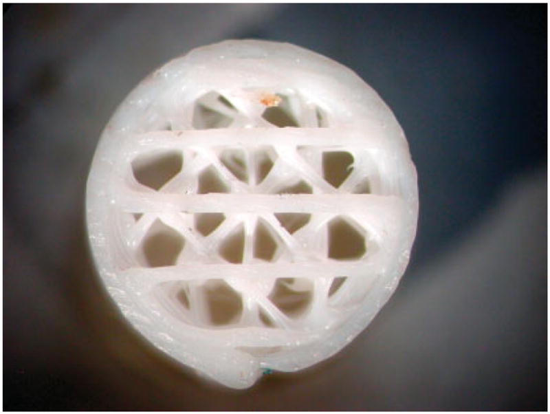
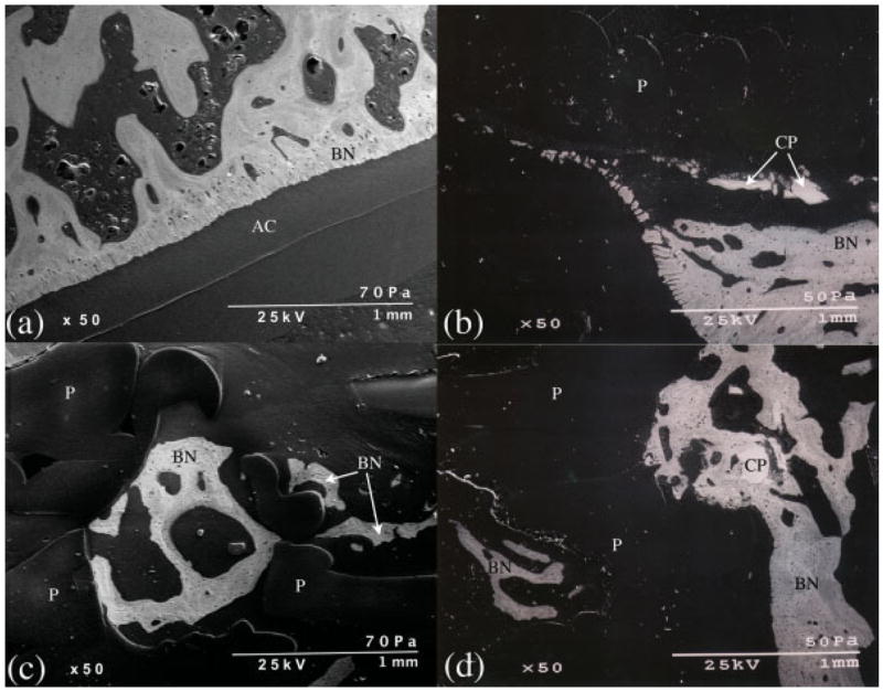
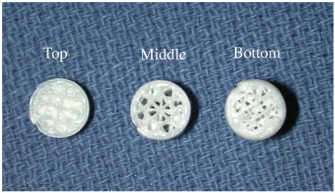
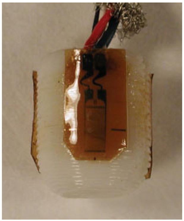
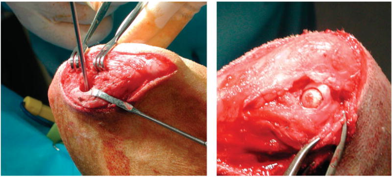
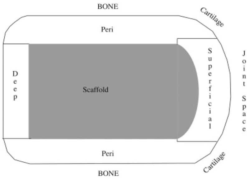
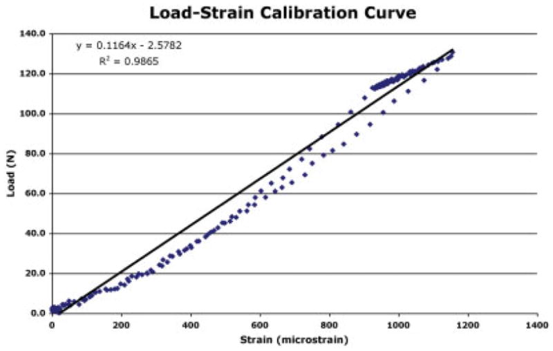
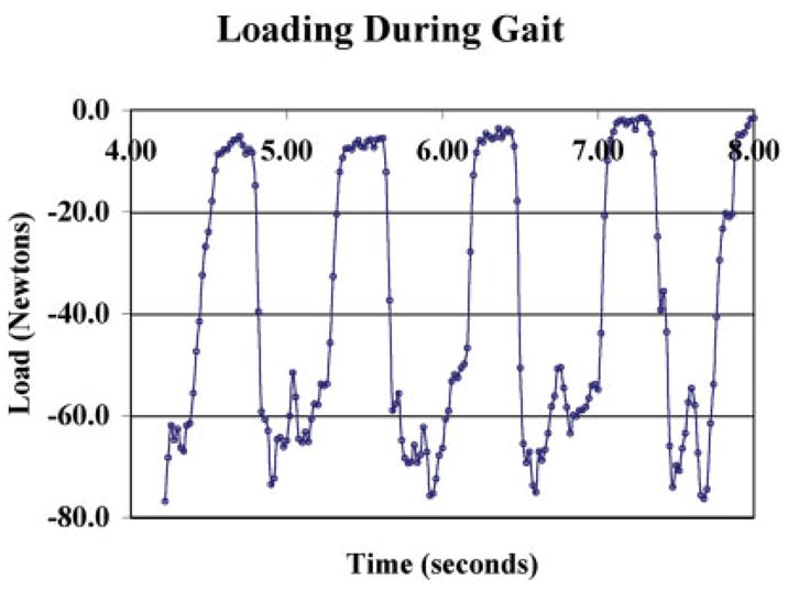
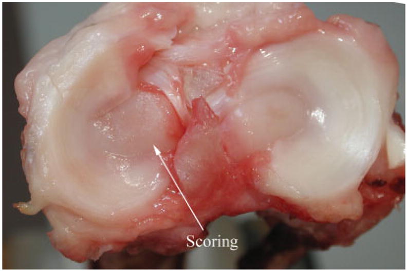
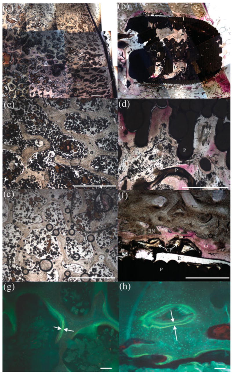
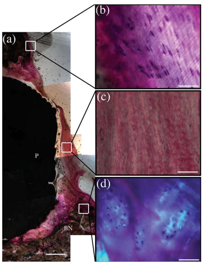
No technique has been consistently successful in the repair of large focal defects in cartilage, particularly in older patients. Tissue-engineered cartilage grown on synthetic scaffolds with appropriate mechanical properties will provide an implant, which could be used to treat this problem. A means of monitoring loads and pressures acting on cartilage, at the defect site, will provide information needed to understand integration and survival of engineered tissues. It will also provide a means of evaluating rehabilitation protocols. A "sensate" scaffold with calibrated strain sensors attached to its surface, combined with a subminiature radio transmitter, was developed and utilized to measure loads and pressures during gait. In an animal study utilizing six dogs, peak loads of 120N and peak pressures of 11 MPa were measured during relaxed gait. Ingrowth into the scaffold characterized after 6 months in vivo indicated that it was well anchored and bone formation was continuing. Cartilage tissue formation was noted at the edges of the defect at the joint-scaffold interfaces. This suggested that native cartilage integration in future formulations of this scaffold configured with engineered cartilage will be a possibility.

摘要

在修复软骨的大面积局灶性缺损方面,尚无一种技术能始终取得成功,尤其是在老年患者中。在具有适当机械性能的合成支架上生长的组织工程软骨将提供一种植入物,可用于治疗这一问题。一种监测作用于缺损部位软骨的负荷和压力的方法,将提供了解工程组织整合和存活所需的信息。它还将提供一种评估康复方案的方法。开发了一种“传感”支架,其表面附着有校准应变传感器,并结合一个超小型无线电发射器,用于测量步态期间的负荷和压力。在一项利用6只狗的动物研究中,在轻松步态期间测量到的峰值负荷为120N,峰值压力为11MPa。体内6个月后对支架内生长情况的表征表明,它固定良好,骨形成仍在继续。在关节-支架界面处的缺损边缘发现了软骨组织形成。这表明,在这种配置有工程软骨的支架的未来配方中,天然软骨整合将是可能的。

https://cdn.ncbi.nlm.nih.gov/pmc/blobs/21e2/2288746/e57e497f8f52/nihms23607f11.jpg
https://cdn.ncbi.nlm.nih.gov/pmc/blobs/21e2/2288746/438948c137dc/nihms23607f1.jpg
https://cdn.ncbi.nlm.nih.gov/pmc/blobs/21e2/2288746/8d5887430d23/nihms23607f2.jpg
https://cdn.ncbi.nlm.nih.gov/pmc/blobs/21e2/2288746/036d4ff19aeb/nihms23607f3.jpg
https://cdn.ncbi.nlm.nih.gov/pmc/blobs/21e2/2288746/5205e19ece6e/nihms23607f4.jpg
https://cdn.ncbi.nlm.nih.gov/pmc/blobs/21e2/2288746/a8b979ae9248/nihms23607f5.jpg
https://cdn.ncbi.nlm.nih.gov/pmc/blobs/21e2/2288746/105130c5c241/nihms23607f6.jpg
https://cdn.ncbi.nlm.nih.gov/pmc/blobs/21e2/2288746/642fbc1a29d0/nihms23607f7.jpg
https://cdn.ncbi.nlm.nih.gov/pmc/blobs/21e2/2288746/f3a901d1c1ac/nihms23607f8.jpg
https://cdn.ncbi.nlm.nih.gov/pmc/blobs/21e2/2288746/a33c476d0bd7/nihms23607f9.jpg
https://cdn.ncbi.nlm.nih.gov/pmc/blobs/21e2/2288746/9cba22c436f0/nihms23607f10.jpg
https://cdn.ncbi.nlm.nih.gov/pmc/blobs/21e2/2288746/e57e497f8f52/nihms23607f11.jpg