• 文献检索
  • 文档翻译
  • 深度研究
  • 学术资讯
  • Suppr Zotero 插件Zotero 插件
  • 邀请有礼
  • 套餐&价格
  • 历史记录
应用&插件
Suppr Zotero 插件Zotero 插件浏览器插件Mac 客户端Windows 客户端微信小程序
定价
高级版会员购买积分包购买API积分包
服务
文献检索文档翻译深度研究API 文档MCP 服务
关于我们
关于 Suppr公司介绍联系我们用户协议隐私条款
关注我们

Suppr 超能文献

核心技术专利:CN118964589B侵权必究
粤ICP备2023148730 号-1Suppr @ 2026

文献检索

告别复杂PubMed语法,用中文像聊天一样搜索,搜遍4000万医学文献。AI智能推荐,让科研检索更轻松。

立即免费搜索

文件翻译

保留排版,准确专业,支持PDF/Word/PPT等文件格式,支持 12+语言互译。

免费翻译文档

深度研究

AI帮你快速写综述,25分钟生成高质量综述,智能提取关键信息,辅助科研写作。

立即免费体验

相似文献

1
Time course of substance P expression in dorsal root ganglia following complete spinal nerve transection.完全性脊神经横断后背根神经节中P物质表达的时间进程。
J Comp Neurol. 2006 Jul 1;497(1):78-87. doi: 10.1002/cne.20981.
2
ATF3 expression in L4 dorsal root ganglion neurons after L5 spinal nerve transection.L5脊神经横断后L4背根神经节神经元中ATF3的表达
Eur J Neurosci. 2006 Jan;23(2):365-73. doi: 10.1111/j.1460-9568.2005.04568.x.
3
Restoration of substance P and calcitonin gene-related peptide in dorsal root ganglia and dorsal horn after neonatal sciatic nerve lesion.新生大鼠坐骨神经损伤后背根神经节和脊髓背角中P物质和降钙素基因相关肽的恢复
J Comp Neurol. 1993 Aug 15;334(3):370-84. doi: 10.1002/cne.903340304.
4
NGF rescues substance P expression but not neurofilament or tubulin gene expression in axotomized sensory neurons.神经生长因子可挽救切断轴突的感觉神经元中P物质的表达,但不能挽救神经丝或微管蛋白基因的表达。
J Neurosci. 1991 Feb;11(2):543-52. doi: 10.1523/JNEUROSCI.11-02-00543.1991.
5
Increase of preprotachykinin mRNA and substance P immunoreactivity in spared dorsal root ganglion neurons following partial sciatic nerve injury.部分坐骨神经损伤后备用背根神经节神经元中前速激肽原mRNA和P物质免疫反应性增加。
Eur J Neurosci. 1998 Jul;10(7):2388-99. doi: 10.1046/j.1460-9568.1998.00249.x.
6
L5 Spinal Nerve Axotomy Induces Distinct Electrophysiological Changes in Axotomized L5- and Adjacent L4-Dorsal Root Ganglion Neurons in Rats .L5 脊神经切断术诱导大鼠损伤的 L5 和相邻 L4 背根神经节神经元产生明显的电生理变化。
J Neurotrauma. 2021 Feb;38(3):330-341. doi: 10.1089/neu.2020.7264. Epub 2020 Oct 27.
7
Change in mRNAs for neuropeptides and the GABA(A) receptor in dorsal root ganglion neurons in a rat experimental neuropathic pain model.大鼠实验性神经病理性疼痛模型中背根神经节神经元神经肽和GABA(A)受体的mRNA变化
Pain. 1998 Oct;78(1):13-26. doi: 10.1016/S0304-3959(98)00111-0.
8
Differential expression of galanin immunoreactivities in the primary sensory neurons following partial and complete sciatic nerve injuries.坐骨神经部分和完全损伤后初级感觉神经元中甘丙肽免疫反应性的差异表达。
Neuroscience. 1997 Aug;79(4):1183-95. doi: 10.1016/s0306-4522(97)00088-2.
9
Expression of auxiliary beta subunits of sodium channels in primary afferent neurons and the effect of nerve injury.初级传入神经元中钠通道辅助β亚基的表达及神经损伤的影响
Neuroscience. 2003;121(2):441-50. doi: 10.1016/s0306-4522(03)00432-9.
10
Brain-derived neurotrophic factor increases in the uninjured dorsal root ganglion neurons in selective spinal nerve ligation model.在选择性脊神经结扎模型中,脑源性神经营养因子在未损伤的背根神经节神经元中增加。
J Neurosci. 2001 Jul 1;21(13):4891-900. doi: 10.1523/JNEUROSCI.21-13-04891.2001.

引用本文的文献

1
Spinal neuromodulation using ultra low frequency waveform inhibits sensory signaling to the thalamus and preferentially reduces aberrant firing of thalamic neurons in a model of neuropathic pain.在神经性疼痛模型中,使用超低频波形的脊髓神经调节可抑制向丘脑的感觉信号传导,并优先减少丘脑神经元的异常放电。
Front Neurosci. 2025 Jan 17;18:1512950. doi: 10.3389/fnins.2024.1512950. eCollection 2024.
2
Assessment of corneal nerve regeneration after axotomy in a compartmentalized microfluidic chip model with automated 3D high resolution live-imaging.在具有自动三维高分辨率实时成像的分隔微流控芯片模型中评估轴突切断术后的角膜神经再生。
Front Cell Neurosci. 2024 Jul 15;18:1417653. doi: 10.3389/fncel.2024.1417653. eCollection 2024.
3
Multicenter assessment of quantitative sensory testing (QST) for the detection of neuropathic-like pain responses using the topical capsaicin model.使用局部辣椒素模型对定量感觉测试(QST)检测神经性疼痛样反应的多中心评估。
Can J Pain. 2018 Oct 23;2(1):266-279. doi: 10.1080/24740527.2018.1525682. eCollection 2018.
4
Prior Acoustic Trauma Alters Type II Afferent Activity in the Mouse Cochlea.先前的声创伤改变了小鼠耳蜗中的 II 型传入活动。
eNeuro. 2021 Nov 11;8(6). doi: 10.1523/ENEURO.0383-21.2021. Print 2021 Nov-Dec.
5
Revisiting PNS Plasticity: How Uninjured Sensory Afferents Promote Neuropathic Pain.重新审视周围神经系统可塑性:未受损的感觉传入神经如何引发神经性疼痛。
Front Cell Neurosci. 2020 Dec 10;14:612982. doi: 10.3389/fncel.2020.612982. eCollection 2020.
6
John J. Bonica Award Lecture: Peripheral neuronal hyperexcitability: the "low-hanging" target for safe therapeutic strategies in neuropathic pain.约翰·J·博尼卡奖演讲:周围神经元兴奋性过高:神经病理性疼痛安全治疗策略的“低挂果实”。
Pain. 2020 Sep;161 Suppl 1(Suppl 1):S14-S26. doi: 10.1097/j.pain.0000000000001838.
7
Contribution of DNMT1 to Neuropathic Pain Genesis Partially through Epigenetically Repressing in Primary Afferent Neurons.DNMT1 对神经病理性疼痛发生的贡献部分是通过表观遗传抑制初级传入神经元中的 实现的。
J Neurosci. 2019 Aug 14;39(33):6595-6607. doi: 10.1523/JNEUROSCI.0695-19.2019. Epub 2019 Jun 10.
8
Subclinical lipopolysaccharide from Salmonella Enteritidis induces neuropeptide dysregulation in the spinal cord and the dorsal root ganglia.肠炎沙门氏菌亚临床内毒素诱导脊髓和背根神经节神经肽失调。
BMC Neurosci. 2019 Apr 25;20(1):18. doi: 10.1186/s12868-019-0502-z.
9
Role of dorsal root ganglion K2p1.1 in peripheral nerve injury-induced neuropathic pain.背根神经节 K2p1.1 在周围神经损伤诱导的神经性疼痛中的作用。
Mol Pain. 2017 Jan;13:1744806917701135. doi: 10.1177/1744806917701135.
10
DNA methyltransferase DNMT3a contributes to neuropathic pain by repressing Kcna2 in primary afferent neurons.DNA 甲基转移酶 DNMT3a 通过抑制初级传入神经元中的 Kcna2 促进神经病理性疼痛。
Nat Commun. 2017 Mar 8;8:14712. doi: 10.1038/ncomms14712.

本文引用的文献

1
Allergen-induced substance P synthesis in large-diameter sensory neurons innervating the lungs.变应原诱导支配肺部的大直径感觉神经元合成P物质。
J Allergy Clin Immunol. 2005 Aug;116(2):325-31. doi: 10.1016/j.jaci.2005.04.005.
2
Nociceptive transmitter release in the dorsal spinal cord by capsaicin-sensitive fibers after noxious gastric stimulation.伤害性胃刺激后,辣椒素敏感纤维在脊髓背角释放伤害性递质。
Brain Res. 2005 Mar 28;1039(1-2):108-15. doi: 10.1016/j.brainres.2005.01.050.
3
Ectopic discharges from injured nerve fibers are highly correlated with tactile allodynia only in early, but not late, stage in rats with spinal nerve ligation.在脊髓神经结扎的大鼠中,受损神经纤维的异位放电仅在早期而非晚期与触觉异常性疼痛高度相关。
Exp Neurol. 2005 Jan;191(1):128-36. doi: 10.1016/j.expneurol.2004.09.008.
4
Partial sciatic nerve transection causes redistribution of pain-related peptides and lowers withdrawal threshold.坐骨神经部分横断会导致疼痛相关肽的重新分布,并降低撤离阈值。
Exp Neurol. 2004 Aug;188(2):444-51. doi: 10.1016/j.expneurol.2004.04.018.
5
Effects of spinal nerve ligation on immunohistochemically identified neurons in the L4 and L5 dorsal root ganglia of the rat.脊髓神经结扎对大鼠L4和L5背根神经节中免疫组化鉴定神经元的影响。
J Comp Neurol. 2004 Aug 2;475(4):575-89. doi: 10.1002/cne.20209.
6
Co-localization of endomorphin-2 and substance P in primary afferent nociceptors and effects of injury: a light and electron microscopic study in the rat.内吗啡肽-2与P物质在初级传入伤害性感受器中的共定位及损伤效应:大鼠的光镜和电镜研究
Eur J Neurosci. 2004 Apr;19(7):1789-99. doi: 10.1111/j.1460-9568.2004.03284.x.
7
Characterization of wide dynamic range neurons in the deep dorsal horn of the spinal cord in preprotachykinin-a null mice in vivo.体内前速激肽原-a基因敲除小鼠脊髓背角深层宽动态范围神经元的特征研究
J Neurophysiol. 2004 May;91(5):1945-54. doi: 10.1152/jn.00945.2003. Epub 2004 Jan 7.
8
Retrograde transport of neurotrophins: fact and function.神经营养因子的逆向运输:事实与功能
J Neurobiol. 2004 Feb 5;58(2):217-29. doi: 10.1002/neu.10322.
9
Pathology of lumbar nerve root compression. Part 2: morphological and immunohistochemical changes of dorsal root ganglion.腰神经根受压的病理学。第2部分:背根神经节的形态学和免疫组化变化
J Orthop Res. 2004 Jan;22(1):180-8. doi: 10.1016/S0736-0266(03)00132-3.
10
Paw withdrawal thresholds and persistent hindlimb flexion in experimental mononeuropathies.
J Pain. 2003 May;4(4):222-30. doi: 10.1016/s1526-5900(03)00619-9.

完全性脊神经横断后背根神经节中P物质表达的时间进程。

Time course of substance P expression in dorsal root ganglia following complete spinal nerve transection.

作者信息

Weissner Wendy, Winterson Barbara J, Stuart-Tilley Alan, Devor Marshall, Bove Geoffrey M

机构信息

Department of Anesthesia and Critical Care, Beth Israel Deaconess Medical Center and Harvard Medical School, Boston, Massachusetts 02215, USA.

出版信息

J Comp Neurol. 2006 Jul 1;497(1):78-87. doi: 10.1002/cne.20981.

DOI:10.1002/cne.20981
PMID:16680762
原文链接:https://pmc.ncbi.nlm.nih.gov/articles/PMC2571959/
Abstract

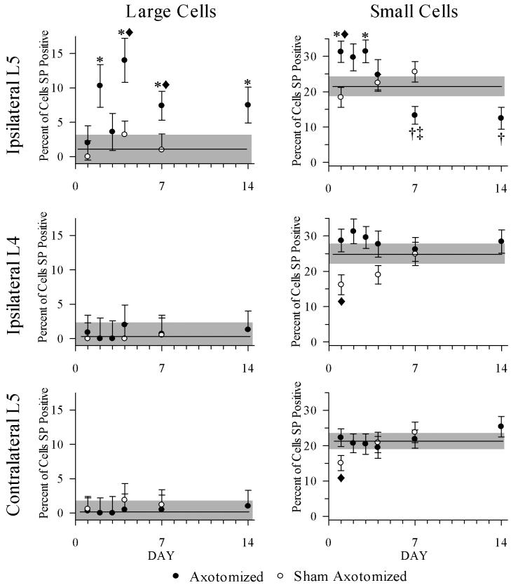
Recent evidence suggests that substance P (SP) is up-regulated in primary sensory neurons following axotomy and that this change occurs in larger neurons that do not usually produce SP. If this is so, then the up-regulation may allow normally neighboring, uninjured, and nonnociceptive dorsal root ganglion (DRG) neurons to become effective in activating pain pathways. By using immunohistochemistry, we performed a unilateral L5 spinal nerve transection on male Wistar rats and measured SP expression in ipsilateral L4 and L5 DRGs and contralateral L5 DRGs at 1-14 days postoperatively (dpo) and in control and sham-operated rats. In normal and sham-operated DRGs, SP was detectable almost exclusively in small neurons (< or =800 microm2). After surgery, the mean size of SP-positive neurons from the axotomized L5 ganglia was greater at 2, 4, 7, and 14 dpo. Among large neurons (>800 microm2) from the axotomized L5, the percentage of SP-positive neurons increased at 2, 4, 7, and 14 dpo. Among small neurons from the axotomized L5, the percentage of SP-positive neurons was increased at 1 and 3 dpo but was decreased at 7 and 14 dpo. Thus, SP expression is affected by axonal damage, and the time course of the expression is different between large and small DRG neurons. These data support a role for SP-producing, large DRG neurons in persistent sensory changes resulting from nerve injury.

摘要

最近的证据表明,在轴突切断后,初级感觉神经元中的P物质(SP)上调,并且这种变化发生在通常不产生SP的较大神经元中。如果是这样,那么这种上调可能会使通常相邻的、未受损的和非伤害性的背根神经节(DRG)神经元有效地激活疼痛通路。通过免疫组织化学方法,我们对雄性Wistar大鼠进行了单侧L5脊神经切断术,并在术后1至14天(dpo)测量了同侧L4和L5 DRG以及对侧L5 DRG中SP的表达,并与对照和假手术大鼠进行了比较。在正常和假手术的DRG中,几乎仅在小神经元(≤800平方微米)中可检测到SP。手术后,在2、4、7和14 dpo时,来自切断轴突的L5神经节的SP阳性神经元的平均大小更大。在切断轴突的L5的大神经元(> 800平方微米)中,SP阳性神经元的百分比在2、4、7和14 dpo时增加。在切断轴突的L5的小神经元中,SP阳性神经元的百分比在1和3 dpo时增加,但在7和14 dpo时降低。因此,SP表达受轴突损伤影响,并且大小不同的DRG神经元之间表达的时间进程也不同。这些数据支持产生SP的大DRG神经元在神经损伤导致的持续性感觉变化中起作用。

https://cdn.ncbi.nlm.nih.gov/pmc/blobs/3929/2571959/a8bea878c444/nihms-70197-f0005.jpg
https://cdn.ncbi.nlm.nih.gov/pmc/blobs/3929/2571959/5abc37d61963/nihms-70197-f0001.jpg
https://cdn.ncbi.nlm.nih.gov/pmc/blobs/3929/2571959/ffb9521fe0e3/nihms-70197-f0002.jpg
https://cdn.ncbi.nlm.nih.gov/pmc/blobs/3929/2571959/ece93e9b5e80/nihms-70197-f0003.jpg
https://cdn.ncbi.nlm.nih.gov/pmc/blobs/3929/2571959/030e3febd299/nihms-70197-f0004.jpg
https://cdn.ncbi.nlm.nih.gov/pmc/blobs/3929/2571959/a8bea878c444/nihms-70197-f0005.jpg