Ala-Laurila Petri, Kolesnikov Alexander V, Crouch Rosalie K, Tsina Efthymia, Shukolyukov Sergey A, Govardovskii Victor I, Koutalos Yiannis, Wiggert Barbara, Estevez Maureen E, Cornwall M Carter
Department of Physiology and Biophysics, Boston University School of Medicine, Boston, MA 02118, USA.
J Gen Physiol. 2006 Aug;128(2):153-69. doi: 10.1085/jgp.200609557. Epub 2006 Jul 17.
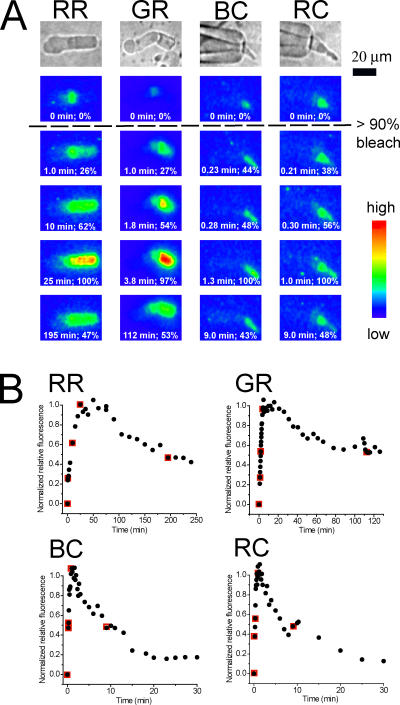
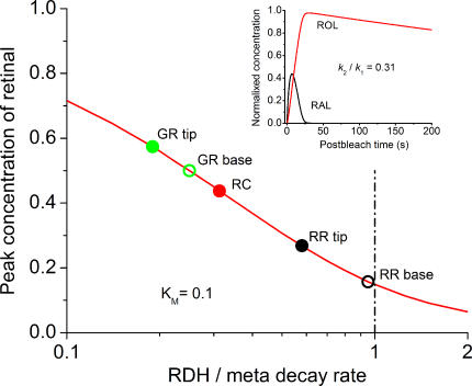
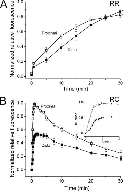
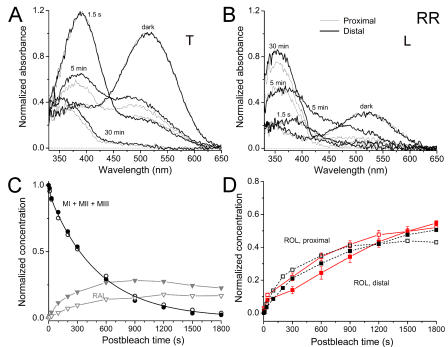
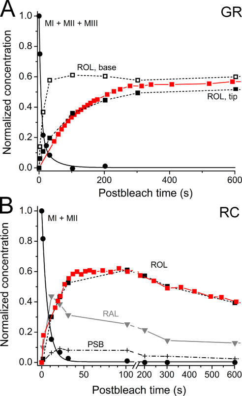
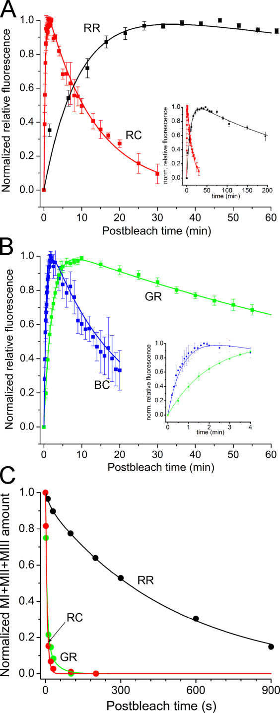
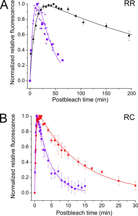
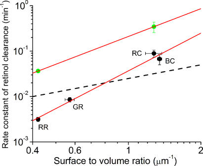
The visual cycle is a chain of biochemical reactions that regenerate visual pigment following exposure to light. Initial steps, the liberation of all-trans retinal and its reduction to all-trans retinol by retinol dehydrogenase (RDH), take place in photoreceptors. We performed comparative microspectrophotometric and microfluorometric measurements on a variety of rod and cone photoreceptors isolated from salamander retinae to correlate the rates of photoproduct decay and retinol production. Metapigment decay rate was spatially uniform within outer segments and 50-70 times faster in the cells that contained cone-type pigment (SWS2 and M/LWS) compared to cells with rod-type pigment (RH1). Retinol production rate was strongly position dependent, fastest at the base of outer segments. Retinol production rate was 10-40 times faster in cones with cone pigments (SWS2 and M/LWS) than in the basal OS of rods containing rod pigment (RH1). Production rate was approximately five times faster in rods containing cone pigment (SWS2) than the rate in basal OS of rods containing the rod pigment (RH1). We show that retinol production is defined either by metapigment decay rate or RDH reaction rate, depending on cell type or outer segment region, whereas retinol removal is defined by the surface-to-volume ratio of the outer segment and the availability of retinoid binding protein (IRBP). The more rapid rates of retinol production in cones compared to rods are consistent with the more rapid operation of the visual cycle in these cells.
视觉循环是一系列生化反应,在光照后使视觉色素再生。初始步骤,即全反式视黄醛的释放及其被视黄醇脱氢酶(RDH)还原为全反式视黄醇,发生在光感受器中。我们对从蝾螈视网膜分离出的多种视杆和视锥光感受器进行了比较显微分光光度法和显微荧光测定,以关联光产物衰变速率和视黄醇生成速率。间色素衰变速率在外段内空间上是均匀的,与含有视杆型色素(RH1)的细胞相比,含有视锥型色素(SWS2和M/LWS)的细胞中的衰变速率快50 - 70倍。视黄醇生成速率强烈依赖于位置,在外段基部最快。含有视锥色素(SWS2和M/LWS)的视锥细胞中的视黄醇生成速率比含有视杆色素(RH1)的视杆细胞基部外段快10 - 40倍。含有视锥色素(SWS2)的视杆细胞中的生成速率比含有视杆色素(RH1)的视杆细胞基部外段的速率快约五倍。我们表明,视黄醇生成由间色素衰变速率或RDH反应速率决定,这取决于细胞类型或外段区域,而视黄醇的去除由外段的表面积与体积之比以及类视黄醇结合蛋白(IRBP)的可用性决定。与视杆细胞相比,视锥细胞中视黄醇生成速率更快,这与这些细胞中视觉循环的更快运行一致。