• 文献检索
  • 文档翻译
  • 深度研究
  • 学术资讯
  • Suppr Zotero 插件Zotero 插件
  • 邀请有礼
  • 套餐&价格
  • 历史记录
应用&插件
Suppr Zotero 插件Zotero 插件浏览器插件Mac 客户端Windows 客户端微信小程序
定价
高级版会员购买积分包购买API积分包
服务
文献检索文档翻译深度研究API 文档MCP 服务
关于我们
关于 Suppr公司介绍联系我们用户协议隐私条款
关注我们

Suppr 超能文献

核心技术专利:CN118964589B侵权必究
粤ICP备2023148730 号-1Suppr @ 2026

文献检索

告别复杂PubMed语法,用中文像聊天一样搜索,搜遍4000万医学文献。AI智能推荐,让科研检索更轻松。

立即免费搜索

文件翻译

保留排版,准确专业,支持PDF/Word/PPT等文件格式,支持 12+语言互译。

免费翻译文档

深度研究

AI帮你快速写综述,25分钟生成高质量综述,智能提取关键信息,辅助科研写作。

立即免费体验

扩散加权磁共振成像及其在癌症中的应用。

Diffusion-weighted magnetic resonance imaging and its application to cancer.

作者信息

Charles-Edwards Elizabeth M, deSouza Nandita M

机构信息

Cancer Research UK Clinical Magnetic Resonance Research Group, Institute of Cancer Research and Royal Marsden NHS Foundation Trust, Sutton, Surrey, UK.

出版信息

Cancer Imaging. 2006 Sep 13;6(1):135-43. doi: 10.1102/1470-7330.2006.0021.

DOI:10.1102/1470-7330.2006.0021
PMID:17015238
原文链接:https://pmc.ncbi.nlm.nih.gov/articles/PMC1693785/
Abstract

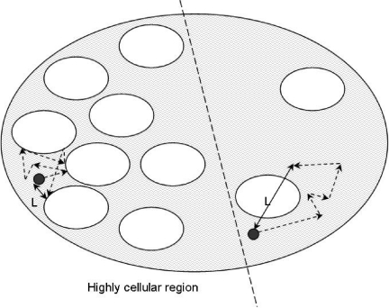
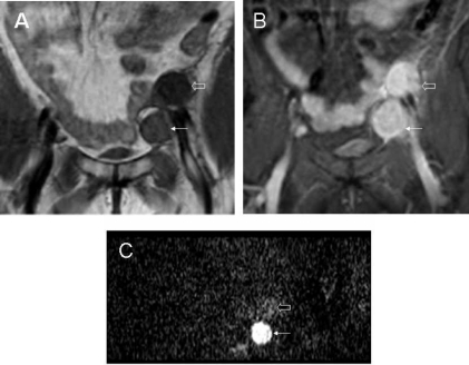
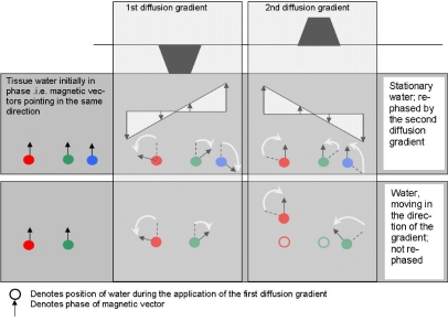
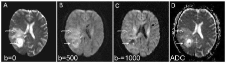
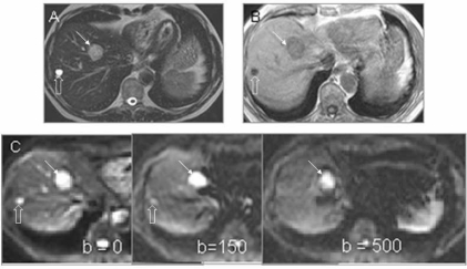
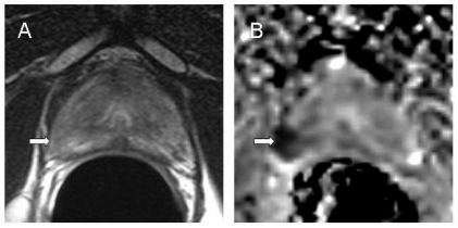
Diffusion-weighted magnetic resonance imaging (DW-MRI) provides image contrast through measurement of the diffusion properties of water within tissues. Application of diffusion sensitising gradients to the MR pulse sequence allows water molecular displacement over distances of 1-20 microm to be recognised. Diffusion can be predominantly unidirectional (anisotropic) or not (isotropic). Combining images obtained with different amounts of diffusion weighting provides an apparent diffusion coefficient (ADC) map. In cancer imaging DW-MRI has been used to distinguish brain tumours from peritumoural oedema. It is also increasingly exploited to differentiate benign and malignant lesions in liver, breast and prostate where increased cellularity of malignant lesions restricts water motion in a reduced extracellular space. It is proving valuable in monitoring treatment where changes due to cell swelling and apoptosis are measurable as changes in ADC at an earlier stage than subsequent conventional radiological response indicators.

摘要

扩散加权磁共振成像(DW-MRI)通过测量组织内水的扩散特性来提供图像对比度。在MR脉冲序列中应用扩散敏感梯度可识别水分子在1-20微米距离上的位移。扩散可以主要是单向的(各向异性)或不是(各向同性)。将用不同扩散加权量获得的图像相结合可提供表观扩散系数(ADC)图。在癌症成像中,DW-MRI已被用于区分脑肿瘤与瘤周水肿。它也越来越多地被用于区分肝脏、乳腺和前列腺中的良性和恶性病变,其中恶性病变细胞增多限制了细胞外空间减少时的水运动。事实证明,它在监测治疗方面很有价值,因为与随后的传统放射学反应指标相比,细胞肿胀和凋亡引起的变化在更早阶段就可作为ADC的变化进行测量。

https://cdn.ncbi.nlm.nih.gov/pmc/blobs/10e5/1693785/da1325d1d8c1/ci06013506.jpg
https://cdn.ncbi.nlm.nih.gov/pmc/blobs/10e5/1693785/bb3e450c618d/ci06013501.jpg
https://cdn.ncbi.nlm.nih.gov/pmc/blobs/10e5/1693785/dabd22467fa6/ci06013502.jpg
https://cdn.ncbi.nlm.nih.gov/pmc/blobs/10e5/1693785/eb1e64f6f4a3/ci06013503.jpg
https://cdn.ncbi.nlm.nih.gov/pmc/blobs/10e5/1693785/1b579f75cae1/ci06013504.jpg
https://cdn.ncbi.nlm.nih.gov/pmc/blobs/10e5/1693785/7a537f06751a/ci06013505.jpg
https://cdn.ncbi.nlm.nih.gov/pmc/blobs/10e5/1693785/da1325d1d8c1/ci06013506.jpg

相似文献

1
Diffusion-weighted magnetic resonance imaging and its application to cancer.扩散加权磁共振成像及其在癌症中的应用。
Cancer Imaging. 2006 Sep 13;6(1):135-43. doi: 10.1102/1470-7330.2006.0021.
2
Magnetic resonance diffusion-weighted imaging: extraneurological applications.磁共振扩散加权成像:神经外应用
Radiol Med. 2006 Apr;111(3):392-419. doi: 10.1007/s11547-006-0037-0. Epub 2006 Apr 11.
3
Diffusion weighted imaging in breast MRI: comparison of two different pulse sequences.乳腺MRI中的扩散加权成像:两种不同脉冲序列的比较
Acad Radiol. 2007 Sep;14(9):1077-83. doi: 10.1016/j.acra.2007.06.006.
4
Assessing extracranial tumors using diffusion-weighted whole-body MRI.使用扩散加权全身 MRI 评估颅外肿瘤。
Z Med Phys. 2011 May;21(2):79-90. doi: 10.1016/j.zemedi.2010.06.004. Epub 2010 Oct 2.
5
In vivo measurement of the apparent diffusion coefficient in normal and malignant prostatic tissue using thin-slice echo-planar imaging.使用薄层回波平面成像对正常和恶性前列腺组织中的表观扩散系数进行体内测量。
Radiol Med. 2006 Dec;111(8):1124-33. doi: 10.1007/s11547-006-0110-8. Epub 2006 Dec 20.
6
Usefulness of diffusion-weighted imaging and dynamic contrast-enhanced magnetic resonance imaging in the diagnosis of prostate transition-zone cancer.扩散加权成像和动态对比增强磁共振成像在前列腺移行区癌诊断中的应用价值
Acta Radiol. 2008 Dec;49(10):1207-13. doi: 10.1080/02841850802508959.
7
Diffusion-weighted magnetic resonance imaging of breast lesions: first experiences at 3 T.乳腺病变的扩散加权磁共振成像:3T场强下的初步经验
J Comput Assist Tomogr. 2009 Jan-Feb;33(1):63-9. doi: 10.1097/RCT.0b013e318165dc6b.
8
Role of diffusion-weighted magnetic resonance imaging in differentiation between the viable and necrotic parts of head and neck tumors.扩散加权磁共振成像在头颈部肿瘤存活部分与坏死部分鉴别中的作用。
Acta Radiol. 2008 Apr;49(3):364-70. doi: 10.1080/02841850701777390.
9
Diffusion-weighted MR imaging for whole body metastatic disease and lymphadenopathy.用于全身转移性疾病和淋巴结病的扩散加权磁共振成像。
Magn Reson Imaging Clin N Am. 2009 May;17(2):245-61. doi: 10.1016/j.mric.2009.01.006.
10
Tissue characterization of head and neck lesions using diffusion-weighted MR imaging with SPLICE.使用具有SPLICE的扩散加权磁共振成像对头颈部病变进行组织特征分析。
Eur J Radiol. 2009 Feb;69(2):260-8. doi: 10.1016/j.ejrad.2007.10.008. Epub 2007 Nov 26.

引用本文的文献

1
Local response function estimation in spherical deconvolution for comprehensive tissue representation using diffusion MRI.利用扩散磁共振成像在球形反褶积中进行局部响应函数估计以实现全面的组织表征
Imaging Neurosci (Camb). 2025 Aug 1;3. doi: 10.1162/IMAG.a.95. eCollection 2025.
2
The use of diffusion-weighted magnetic resonance imaging and parametric response mapping for disease outcome prediction in nasopharyngeal carcinoma.利用扩散加权磁共振成像和参数反应映射预测鼻咽癌的疾病转归
BMC Med Imaging. 2025 Jul 31;25(1):308. doi: 10.1186/s12880-025-01847-2.
3
Multiparametric MRI in Diagnosis of Parotid Gland Tumor: An Observational Study in 3-T MRI.

本文引用的文献

1
Diffusion-weighted and perfusion MR imaging for brain tumor characterization and assessment of treatment response.用于脑肿瘤特征描述及治疗反应评估的扩散加权磁共振成像和灌注磁共振成像
Radiology. 2006 Jun;239(3):632-49. doi: 10.1148/radiol.2393042031.
2
Colorectal hepatic metastases: quantitative measurements using single-shot echo-planar diffusion-weighted MR imaging.结直肠癌肝转移:使用单次激发回波平面扩散加权磁共振成像进行定量测量
Eur Radiol. 2006 Sep;16(9):1898-905. doi: 10.1007/s00330-006-0201-x. Epub 2006 May 12.
3
Neoadjuvant chemotherapy in breast cancer: early response prediction with quantitative MR imaging and spectroscopy.
多参数磁共振成像在腮腺肿瘤诊断中的应用:一项3-T磁共振成像的观察性研究
Indian J Radiol Imaging. 2024 Dec 17;35(3):402-410. doi: 10.1055/s-0044-1800861. eCollection 2025 Jul.
4
Utility of multiparametric MRI including T1/T2 mapping and IVIM/diffusion imaging for the evaluation of non-obstructive azoospermia.包括T1/T2映射和体素内不相干运动/扩散成像在内的多参数磁共振成像在评估非梗阻性无精子症中的应用。
MAGMA. 2025 Jun 14. doi: 10.1007/s10334-025-01267-x.
5
Differential tractography: an imaging marker for tissue degeneration in neurodegenerative diseases.差异纤维束成像:神经退行性疾病中组织退化的一种影像学标志物。
Brain Commun. 2025 May 24;7(3):fcaf198. doi: 10.1093/braincomms/fcaf198. eCollection 2025.
6
Tumor ADC value predicts outcome and yields refined prognostication in uterine cervical cancer.肿瘤表观扩散系数(ADC)值可预测子宫颈癌的预后并实现更精准的预后评估。
Cancer Imaging. 2025 Feb 28;25(1):23. doi: 10.1186/s40644-025-00828-6.
7
The Role of Diffusion-Weighted MRI in Correlation with Contrast-Enhanced MRI and Histopathology in the Evaluation of Focal Liver Lesions.扩散加权磁共振成像在局灶性肝病变评估中与对比增强磁共振成像及组织病理学相关性的作用
Cureus. 2024 Oct 11;16(10):e71261. doi: 10.7759/cureus.71261. eCollection 2024 Oct.
8
Differential Tractography: A Biomarker for Neuronal Function in Neurodegenerative Disease.差异纤维束成像:神经退行性疾病中神经元功能的生物标志物
medRxiv. 2024 Aug 26:2024.08.25.24312255. doi: 10.1101/2024.08.25.24312255.
9
Gastrointestinal Stromal Tumors (GISTs) in Pediatric Patients: A Case Report and Literature Review.小儿胃肠道间质瘤(GISTs):一例病例报告及文献综述
Children (Basel). 2024 Aug 26;11(9):1040. doi: 10.3390/children11091040.
10
Multiparametric Whole-Body MRI: A Game Changer in Metastatic Prostate Cancer.多参数全身磁共振成像:转移性前列腺癌治疗的变革者
Cancers (Basel). 2024 Jul 13;16(14):2531. doi: 10.3390/cancers16142531.
乳腺癌的新辅助化疗:利用定量磁共振成像和波谱进行早期反应预测
Br J Cancer. 2006 Feb 13;94(3):427-35. doi: 10.1038/sj.bjc.6602948.
4
[Influence of respiratory motion in body diffusion weighted imaging under free breathing (examination of a moving phantom)].
Nihon Hoshasen Gijutsu Gakkai Zasshi. 2005 Nov 20;61(11):1551-8. doi: 10.6009/jjrt.kj00004010676.
5
Diffusion-weighted imaging of malignant breast tumors: the usefulness of apparent diffusion coefficient (ADC) value and ADC map for the detection of malignant breast tumors and evaluation of cancer extension.乳腺恶性肿瘤的扩散加权成像:表观扩散系数(ADC)值及ADC图在乳腺恶性肿瘤检测及癌灶范围评估中的应用价值
J Comput Assist Tomogr. 2005 Sep-Oct;29(5):644-9. doi: 10.1097/01.rct.0000171913.74086.1b.
6
Diffusion-weighted imaging of breast cancer with the sensitivity encoding technique: analysis of the apparent diffusion coefficient value.采用敏感性编码技术的乳腺癌扩散加权成像:表观扩散系数值分析
Magn Reson Med Sci. 2004 Jul 15;3(2):79-85. doi: 10.2463/mrms.3.79.
7
Diffusion-weighted MR imaging in monitoring the effect of a vascular targeting agent on rhabdomyosarcoma in rats.磁共振扩散加权成像监测血管靶向剂对大鼠横纹肌肉瘤的作用
Radiology. 2005 Mar;234(3):756-64. doi: 10.1148/radiol.2343031721.
8
Changes in water mobility measured by diffusion MRI predict response of metastatic breast cancer to chemotherapy.通过扩散磁共振成像测量的水分子扩散运动变化可预测转移性乳腺癌对化疗的反应。
Neoplasia. 2004 Nov-Dec;6(6):831-7. doi: 10.1593/neo.03343.
9
Diffusion weighted whole body imaging with background body signal suppression (DWIBS): technical improvement using free breathing, STIR and high resolution 3D display.背景体部信号抑制扩散加权全身成像(DWIBS):利用自由呼吸、短TI反转恢复序列(STIR)及高分辨率三维显示的技术改进
Radiat Med. 2004 Jul-Aug;22(4):275-82.
10
Endorectal diffusion-weighted imaging in prostate cancer to differentiate malignant and benign peripheral zone tissue.前列腺癌的直肠内扩散加权成像用于鉴别恶性和良性外周带组织。
J Magn Reson Imaging. 2004 Oct;20(4):654-61. doi: 10.1002/jmri.20159.