Eigeliene Natalija, Härkönen Pirkko, Erkkola Risto
Department of Obstetrics and Gynecology, Turku University Central Hospital, 20520 Turku, Finland.
BMC Cancer. 2006 Oct 18;6:246. doi: 10.1186/1471-2407-6-246.
Human breast tissue undergoes phases of proliferation, differentiation and regression regulated by changes of the levels of circulating sex hormones during the menstrual cycle or aging. Ovarian hormones also likely play a key role in the etiology and biology of breast cancer. Reports concerning the proliferative effects of steroid hormones on the normal epithelium of human breast have been conflicting. Some studies have shown that steroid hormones may predispose breast epithelial cells to malignant changes by stimulating their proliferation, which is known to be regulated tightly by stromal cells. The aim of this study was to investigate the effects of 17beta-estradiol and medroxyprogesterone acetate on proliferation, apoptosis, expression of differentiation markers and steroid hormone receptors in breast epithelium using an in vitro model of freshly isolated human breast tissue, in which a proper interaction of breast epithelium and stroma has been maintained.
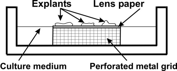
Human breast tissues were obtained from women undergoing surgery for breast tumours. Peritumoral tissues were excised and explants were cultured for 3 weeks in medium supplemented with E2 or MPA or with E2+MPA. Endpoints included histopathological, histomorphometric and immunohistochemical assessment of the breast explants.
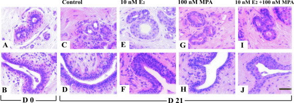
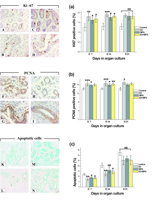
Culture of breast explants for 14 or 21 days with steroid hormones increased proliferative activity and the thickness of acinar and ductal epithelium. E2-treatment led to hyperplastic epithelial morphology, MPA to hypersecretory single-layered epithelium and E2+MPA to multilayered but organised epithelium. The proliferative response to E2 in comparison to control (p < 0.001) was more pronounced than to MPA (p < 0.05) or E2+MPA (p < 0.05) at 7 and 14 days for Ki-67 and PCNA. E2 treatment also decreased the proportion of apoptotic cells after 7 (p < 0.01) and 14 (p < 0.01) days. In addition, the relative number of ERalpha, ERbeta and PR positive epithelial cells was decreased by all hormonal treatments.
Organ culture system provides a model for studying the direct effects of steroid hormones and their analogues on postmenopausal human breast tissue. Addition of E2 or MPA or E2+MPA to breast explants caused characteristic changes in morphology, stimulated epithelial proliferation, lowered apoptosis ratio and decreased the relative number of epithelial cells expressing ERalpha, ERbeta and PR.
在月经周期或衰老过程中,人类乳腺组织会经历增殖、分化和退化阶段,这些阶段受循环性激素水平变化的调节。卵巢激素可能在乳腺癌的病因学和生物学中也起着关键作用。关于甾体激素对人乳腺正常上皮细胞增殖作用的报道一直存在争议。一些研究表明,甾体激素可能通过刺激乳腺上皮细胞增殖,使其易发生恶性变化,而这种增殖已知受基质细胞的严格调控。本研究的目的是使用新鲜分离的人乳腺组织体外模型,研究17β - 雌二醇和醋酸甲羟孕酮对乳腺上皮细胞增殖、凋亡、分化标志物表达和甾体激素受体的影响,该模型能维持乳腺上皮和基质的适当相互作用。
从接受乳腺肿瘤手术的女性身上获取人乳腺组织。切除肿瘤周围组织,将外植体在补充有E2或MPA或E2 + MPA的培养基中培养3周。观察指标包括对乳腺外植体的组织病理学、组织形态计量学和免疫组织化学评估。
用甾体激素培养乳腺外植体14天或21天可增加增殖活性以及腺泡和导管上皮的厚度。E2处理导致上皮形态增生,MPA导致单层上皮分泌亢进,E2 + MPA导致多层但有组织的上皮。在第7天和第14天,与对照相比,Ki - 67和PCNA对E2的增殖反应(p < 0.0(此处原文有误,推测为p < 0.001))比对MPA(p < 0.05)或E2 + MPA(p <0.05)更明显。E2处理在第7天(p < 0.01)和第14天(p < 0.01)也降低了凋亡细胞的比例。此外,所有激素处理均降低了ERα、ERβ和PR阳性上皮细胞的相对数量。
器官培养系统为研究甾体激素及其类似物对绝经后人乳腺组织的直接作用提供了一个模型。向乳腺外植体中添加E2或MPA或E2 + MPA会导致形态学上的特征性变化,刺激上皮细胞增殖,降低凋亡率,并减少表达ERα、ERβ和PR的上皮细胞的相对数量。