• 文献检索
  • 文档翻译
  • 深度研究
  • 学术资讯
  • Suppr Zotero 插件Zotero 插件
  • 邀请有礼
  • 套餐&价格
  • 历史记录
应用&插件
Suppr Zotero 插件Zotero 插件浏览器插件Mac 客户端Windows 客户端微信小程序
定价
高级版会员购买积分包购买API积分包
服务
文献检索文档翻译深度研究API 文档MCP 服务
关于我们
关于 Suppr公司介绍联系我们用户协议隐私条款
关注我们

Suppr 超能文献

核心技术专利:CN118964589B侵权必究
粤ICP备2023148730 号-1Suppr @ 2026

文献检索

告别复杂PubMed语法,用中文像聊天一样搜索,搜遍4000万医学文献。AI智能推荐,让科研检索更轻松。

立即免费搜索

文件翻译

保留排版,准确专业,支持PDF/Word/PPT等文件格式,支持 12+语言互译。

免费翻译文档

深度研究

AI帮你快速写综述,25分钟生成高质量综述,智能提取关键信息,辅助科研写作。

立即免费体验

肿瘤和肉芽组织微血管中内皮细胞、周细胞和基膜标志物的差异表达。

Differential expression of markers for endothelial cells, pericytes, and basal lamina in the microvasculature of tumors and granulation tissue.

作者信息

Schlingemann R O, Rietveld F J, Kwaspen F, van de Kerkhof P C, de Waal R M, Ruiter D J

机构信息

Department of Pathology, Nijmegen University Cancer Center, The Netherlands.

出版信息

Am J Pathol. 1991 Jun;138(6):1335-47.

PMID:1711288
原文链接:https://pmc.ncbi.nlm.nih.gov/articles/PMC1886404/
Abstract

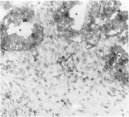
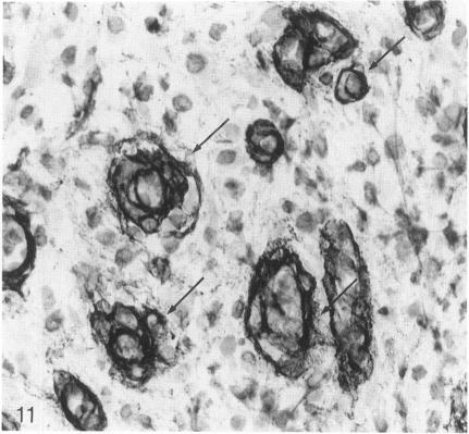
The structure and function of the tumor microvasculature is of great interest for cancer biology, diagnosis, and therapy. The distribution of endothelial cells, pericytes, and basal lamina in tumors is not well documented. In this study, the authors investigated the distribution of markers for these different components in a series of malignant human tumors and in human granulation tissue, both situations with extensive angiogenesis. Their results show a striking heterogeneity in the expression of markers for pericytes and endothelial cells between different tumors, but also within a single tumor lesion. To be able to distinguish between these two adjacent cell types decisively, all marker studies were carried out both on the light and the electron microscopical level and compared with staining results in granulation tissue of cutaneous wounds in healthy volunteers and of decubitus lesions. In granulation tissue of decubitus lesions, well-defined zones with increasing levels of maturation can be delineated. It was found that antibodies recognizing von Willebrand factor often failed to stain the tumor capillaries. Of the pericyte markers, alpha-smooth muscle actin was only locally expressed by pericytes in the tumor vasculature, whereas the high-molecular-weight melanoma-associated antigen, a chondroitin sulfate proteoglycan, stained the microvasculature broadly. Staining of the basal lamina components collagen type IV and laminin was, within the tumor, not restricted to the microvasculature. From their findings the authors conclude that 1) for the visualization of the tumor vasculature, antibodies recognizing endothelial markers, especially monoclonal antibodies PAL-E and BMA 120, are preferable to those recognizing pericytes or basal lamina; 2) within the microvasculature of tumors and granulation tissue, a heterogeneity of expression of endothelial and pericyte markers is observed; 3) during the formation of granulation tissue, all three microvascular components can be demonstrated already in the histologically earliest stage, suggesting not only an involvement of endothelial cells but also of pericytes and basal lamina in the initial steps of angiogenesis in wound healing.

摘要

肿瘤微血管的结构与功能在癌症生物学、诊断及治疗方面备受关注。肿瘤中内皮细胞、周细胞及基膜的分布情况尚无充分记载。在本研究中,作者调查了一系列人类恶性肿瘤及人类肉芽组织(二者均存在广泛血管生成)中这些不同成分标志物的分布。他们的结果显示,不同肿瘤之间以及同一肿瘤病灶内,周细胞和内皮细胞标志物的表达存在显著异质性。为了能够明确区分这两种相邻细胞类型,所有标志物研究均在光学和电子显微镜水平上进行,并与健康志愿者皮肤伤口肉芽组织及褥疮病变的染色结果进行比较。在褥疮病变的肉芽组织中,可以勾勒出成熟度逐渐增加的明确区域。研究发现,识别血管性血友病因子的抗体常常无法使肿瘤毛细血管染色。在周细胞标志物中,α-平滑肌肌动蛋白仅在肿瘤脉管系统的周细胞中局部表达,而高分子量黑素瘤相关抗原(一种硫酸软骨素蛋白聚糖)则广泛地使微血管染色。肿瘤内基膜成分IV型胶原和层粘连蛋白的染色并不局限于微血管。基于这些发现,作者得出以下结论:1)对于肿瘤脉管系统的可视化,识别内皮标志物的抗体,尤其是单克隆抗体PAL-E和BMA 120,优于识别周细胞或基膜的抗体;2)在肿瘤和肉芽组织的微血管系统中,观察到内皮细胞和周细胞标志物表达的异质性;3)在肉芽组织形成过程中,在组织学最早阶段即可显示所有三种微血管成分,这表明在伤口愈合的血管生成初始步骤中,不仅内皮细胞参与其中,周细胞和基膜也参与其中。

https://cdn.ncbi.nlm.nih.gov/pmc/blobs/117d/1886404/3cf762e21f4f/amjpathol00102-0055-b.jpg
https://cdn.ncbi.nlm.nih.gov/pmc/blobs/117d/1886404/e7c5090a491d/amjpathol00102-0049-a.jpg
https://cdn.ncbi.nlm.nih.gov/pmc/blobs/117d/1886404/5cb1f7af157d/amjpathol00102-0050-a.jpg
https://cdn.ncbi.nlm.nih.gov/pmc/blobs/117d/1886404/21b33bd78b35/amjpathol00102-0050-b.jpg
https://cdn.ncbi.nlm.nih.gov/pmc/blobs/117d/1886404/23c52d3390a0/amjpathol00102-0050-c.jpg
https://cdn.ncbi.nlm.nih.gov/pmc/blobs/117d/1886404/9acfb17e388d/amjpathol00102-0051-a.jpg
https://cdn.ncbi.nlm.nih.gov/pmc/blobs/117d/1886404/228f19750596/amjpathol00102-0051-b.jpg
https://cdn.ncbi.nlm.nih.gov/pmc/blobs/117d/1886404/6e8ff11ea1e0/amjpathol00102-0052-a.jpg
https://cdn.ncbi.nlm.nih.gov/pmc/blobs/117d/1886404/65675ae68598/amjpathol00102-0052-b.jpg
https://cdn.ncbi.nlm.nih.gov/pmc/blobs/117d/1886404/99d240eedcdf/amjpathol00102-0053-a.jpg
https://cdn.ncbi.nlm.nih.gov/pmc/blobs/117d/1886404/2d5ec43392ab/amjpathol00102-0053-b.jpg
https://cdn.ncbi.nlm.nih.gov/pmc/blobs/117d/1886404/de9183a6ae6c/amjpathol00102-0053-c.jpg
https://cdn.ncbi.nlm.nih.gov/pmc/blobs/117d/1886404/20ffd2591924/amjpathol00102-0053-d.jpg
https://cdn.ncbi.nlm.nih.gov/pmc/blobs/117d/1886404/267b6efac830/amjpathol00102-0055-a.jpg
https://cdn.ncbi.nlm.nih.gov/pmc/blobs/117d/1886404/3cf762e21f4f/amjpathol00102-0055-b.jpg

相似文献

1
Differential expression of markers for endothelial cells, pericytes, and basal lamina in the microvasculature of tumors and granulation tissue.肿瘤和肉芽组织微血管中内皮细胞、周细胞和基膜标志物的差异表达。
Am J Pathol. 1991 Jun;138(6):1335-47.
2
Angiogenesis in wound healing and tumor metastasis.伤口愈合与肿瘤转移中的血管生成。
Behring Inst Mitt. 1993 Aug(92):258-72.
3
Recruitment of type I collagen producing cells from the microvasculature in vitro.体外从微脉管系统募集产生I型胶原蛋白的细胞。
Exp Cell Res. 1996 Dec 15;229(2):336-49. doi: 10.1006/excr.1996.0379.
4
Expression of matrix metalloprotease-9 in vascular pericytes in human breast cancer.基质金属蛋白酶-9在人乳腺癌血管周细胞中的表达
Lab Invest. 1997 Oct;77(4):345-55.
5
Induction of alpha-smooth muscle actin expression in cultured human brain pericytes by transforming growth factor-beta 1.转化生长因子-β1诱导培养的人脑周细胞中α-平滑肌肌动蛋白表达
Am J Pathol. 1994 Feb;144(2):372-82.
6
Microvascular pericytes express platelet-derived growth factor-beta receptors in human healing wounds and colorectal adenocarcinoma.微血管周细胞在人类愈合伤口和结肠直肠癌中表达血小板衍生生长因子-β受体。
Am J Pathol. 1993 Nov;143(5):1377-88.
7
Differentiation of the pericyte in wound healing: The precursor, the process, and the role of the vascular endothelial cell.周细胞在伤口愈合中的分化:前体、过程及血管内皮细胞的作用
Wound Repair Regen. 2008 May-Jun;16(3):346-55. doi: 10.1111/j.1524-475X.2008.00374.x.
8
Differential expression of alpha-actin mRNA and immunoreactive protein in brain microvascular pericytes and smooth muscle cells.α-肌动蛋白mRNA和免疫反应性蛋白在脑微血管周细胞和平滑肌细胞中的差异表达。
J Neurosci Res. 1994 Nov 1;39(4):430-5. doi: 10.1002/jnr.490390410.
9
Heterogeneity of angiogenesis and blood vessel maturation in human tumors: implications for antiangiogenic tumor therapies.人类肿瘤中血管生成与血管成熟的异质性:对肿瘤抗血管生成治疗的启示
Cancer Res. 2000 Mar 1;60(5):1388-93.
10
Expression of the high molecular weight melanoma-associated antigen by pericytes during angiogenesis in tumors and in healing wounds.肿瘤血管生成和伤口愈合过程中周细胞对高分子量黑素瘤相关抗原的表达。
Am J Pathol. 1990 Jun;136(6):1393-405.

引用本文的文献

1
Optimized enrichment of murine blood-brain barrier vessels with a critical focus on network hierarchy in post-collection analysis.优化小鼠血脑屏障血管的富集,重点关注采集后分析中的网络层次结构。
Sci Rep. 2025 May 6;15(1):15778. doi: 10.1038/s41598-025-99364-3.
2
Development of Immunostaining Protocols for 3D Visualization of Pericytes in Human Retinal Flatmounts.用于人视网膜平铺片周细胞三维可视化的免疫染色方案的开发
J Histochem Cytochem. 2025 Mar-Apr;73(3-4):147-170. doi: 10.1369/00221554251323655. Epub 2025 Mar 17.
3
Evaluation of Anti-Angiogenic Therapy Combined with Immunotherapy and Chemotherapy as a Strategy to Treat Locally Advanced and Metastatic Non-Small-Cell Lung Cancer.

本文引用的文献

1
Enzyme-labeled antibodies: preparation and application for the localization of antigens.酶标抗体:抗原定位的制备与应用
J Histochem Cytochem. 1966 Dec;14(12):929-31. doi: 10.1177/14.12.929.
2
STUDIES ON INFLAMMATION. III. GROWING CAPILLARIES: THEIR STRUCTURE AND PERMEABILITY.炎症研究。III. 生长中的毛细血管:其结构与通透性。
Virchows Arch Pathol Anat Physiol Klin Med. 1963 Nov 8;337:97-141.
3
Retinal vascular patterns. VI. Mural cells of the retinal capillaries.视网膜血管模式。VI. 视网膜毛细血管的壁细胞。
评估抗血管生成疗法联合免疫疗法和化疗作为治疗局部晚期和转移性非小细胞肺癌的策略
Cancers (Basel). 2024 Dec 17;16(24):4207. doi: 10.3390/cancers16244207.
4
New Insights in ATP Synthesis as Therapeutic Target in Cancer and Angiogenic Ocular Diseases.ATP 合成的新见解——作为癌症和血管生成性眼病治疗靶点。
J Histochem Cytochem. 2024 May;72(5):329-352. doi: 10.1369/00221554241249515. Epub 2024 May 11.
5
A tissue engineered 3D printed calcium alkali phosphate bioceramic bone graft enables vascularization and regeneration of critical-size discontinuity bony defects .一种组织工程3D打印钙碱磷酸盐生物陶瓷骨移植体能够实现临界尺寸骨缺损的血管化和再生。
Front Bioeng Biotechnol. 2023 Jun 15;11:1221314. doi: 10.3389/fbioe.2023.1221314. eCollection 2023.
6
PARP inhibition promotes endothelial-like traits in melanoma cells and modulates pericyte coverage dynamics during vasculogenic mimicry.PARP 抑制剂促进黑色素瘤细胞呈现内皮样特征,并调节血管生成拟态过程中的周细胞覆盖动力学。
J Pathol. 2023 Mar;259(3):318-330. doi: 10.1002/path.6043. Epub 2023 Jan 4.
7
Comparison of the Behavior of Perivascular Cells (Pericytes and CD34+ Stromal Cell/Telocytes) in Sprouting and Intussusceptive Angiogenesis.血管周细胞(周细胞和 CD34+基质细胞/间质细胞)在发芽和出芽性血管生成中的行为比较。
Int J Mol Sci. 2022 Aug 12;23(16):9010. doi: 10.3390/ijms23169010.
8
Pericytes in Vascular Development.血管发育中的周细胞
Curr Tissue Microenviron Rep. 2020 Sep;1(3):143-154. doi: 10.1007/s43152-020-00014-9. Epub 2020 Jul 2.
9
Regulation of neovasculogenesis in co-cultures of aortic adventitial fibroblasts and microvascular endothelial cells by cell-cell interactions and TGF-β/ALK5 signaling.细胞间相互作用和TGF-β/ALK5信号通路对主动脉外膜成纤维细胞与微血管内皮细胞共培养中新血管生成的调控
PLoS One. 2020 Dec 28;15(12):e0244243. doi: 10.1371/journal.pone.0244243. eCollection 2020.
10
The role of plasmalemma vesicle-associated protein in pathological breakdown of blood-brain and blood-retinal barriers: potential novel therapeutic target for cerebral edema and diabetic macular edema.质膜小泡相关蛋白在血脑和血视网膜屏障病理破坏中的作用:脑水肿和糖尿病性黄斑水肿潜在的新治疗靶点。
Fluids Barriers CNS. 2018 Sep 20;15(1):24. doi: 10.1186/s12987-018-0109-2.
Arch Ophthalmol. 1963 Apr;69:492-502. doi: 10.1001/archopht.1963.00960040498013.
4
Ultrastructure of the vasculature of central nervous system tumors of childhood.儿童中枢神经系统肿瘤脉管系统的超微结构
Childs Brain. 1980;6(5):242-54. doi: 10.1159/000119910.
5
Endothelial cell proliferation as a novel approach to targeting tumour therapy.内皮细胞增殖作为一种靶向肿瘤治疗的新方法。
Br J Cancer. 1982 Jan;45(1):136-9. doi: 10.1038/bjc.1982.16.
6
Blood vessel fibronectin increases in conjunction with endothelial cell proliferation and capillary ingrowth during wound healing.在伤口愈合过程中,血管纤连蛋白随着内皮细胞增殖和毛细血管向内生长而增加。
J Invest Dermatol. 1982 Nov;79(5):269-76. doi: 10.1111/1523-1747.ep12500076.
7
Growth and vascular structure of human melanoma xenografts.人黑色素瘤异种移植瘤的生长与血管结构
Cell Tissue Kinet. 1984 Jan;17(1):91-101. doi: 10.1111/j.1365-2184.1984.tb00571.x.
8
Factor VIII as a marker of endothelial cells in follicular carcinoma of the thyroid.因子VIII作为甲状腺滤泡癌中内皮细胞的标志物。
J Clin Pathol. 1983 Sep;36(9):1050-4. doi: 10.1136/jcp.36.9.1050.
9
Localization of type IV collagen, laminin, heparan sulfate proteoglycan, and fibronectin to the basal lamina of basement membranes.IV型胶原蛋白、层粘连蛋白、硫酸乙酰肝素蛋白聚糖和纤连蛋白在基底膜的基底层中的定位。
J Cell Biol. 1982 Oct;95(1):340-4. doi: 10.1083/jcb.95.1.340.
10
Factors influencing the neovascularization of experimental tumours.影响实验性肿瘤新生血管形成的因素。
Biorheology. 1984;21(4):493-501. doi: 10.3233/bir-1984-21408.