• 文献检索
  • 文档翻译
  • 深度研究
  • 学术资讯
  • Suppr Zotero 插件Zotero 插件
  • 邀请有礼
  • 套餐&价格
  • 历史记录
应用&插件
Suppr Zotero 插件Zotero 插件浏览器插件Mac 客户端Windows 客户端微信小程序
定价
高级版会员购买积分包购买API积分包
服务
文献检索文档翻译深度研究API 文档MCP 服务
关于我们
关于 Suppr公司介绍联系我们用户协议隐私条款
关注我们

Suppr 超能文献

核心技术专利:CN118964589B侵权必究
粤ICP备2023148730 号-1Suppr @ 2026

文献检索

告别复杂PubMed语法,用中文像聊天一样搜索,搜遍4000万医学文献。AI智能推荐,让科研检索更轻松。

立即免费搜索

文件翻译

保留排版,准确专业,支持PDF/Word/PPT等文件格式,支持 12+语言互译。

免费翻译文档

深度研究

AI帮你快速写综述,25分钟生成高质量综述,智能提取关键信息,辅助科研写作。

立即免费体验

NeuARt II系统:一种基于已发表的神经解剖图谱的神经解剖数据查看工具。

The NeuARt II system: a viewing tool for neuroanatomical data based on published neuroanatomical atlases.

作者信息

Burns Gully A P C, Cheng Wei-Cheng, Thompson Richard H, Swanson Larry W

机构信息

Information Sciences Institute, 4676 Admiralty Way, Marina Del Rey, CA 90292, USA.

出版信息

BMC Bioinformatics. 2006 Dec 13;7:531. doi: 10.1186/1471-2105-7-531.

DOI:10.1186/1471-2105-7-531
PMID:17166289
原文链接:https://pmc.ncbi.nlm.nih.gov/articles/PMC1770939/
Abstract

BACKGROUND

Anatomical studies of neural circuitry describing the basic wiring diagram of the brain produce intrinsically spatial, highly complex data of great value to the neuroscience community. Published neuroanatomical atlases provide a spatial framework for these studies. We have built an informatics framework based on these atlases for the representation of neuroanatomical knowledge. This framework not only captures current methods of anatomical data acquisition and analysis, it allows these studies to be collated, compared and synthesized within a single system.

RESULTS

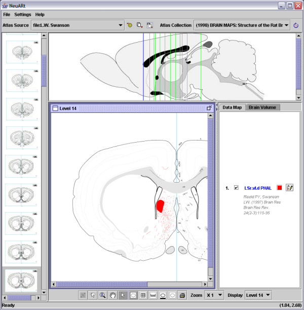
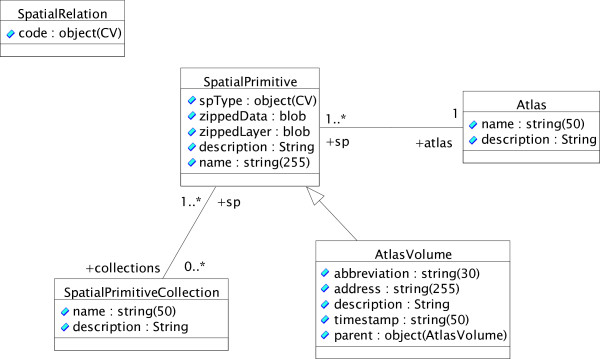
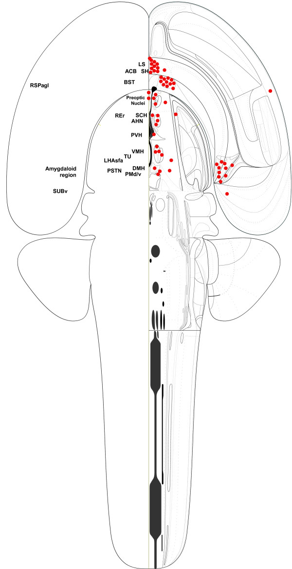
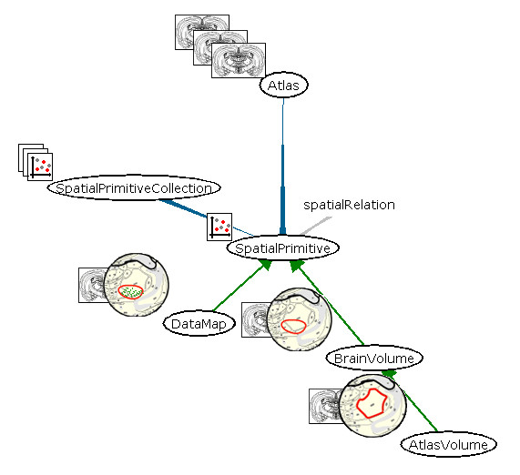
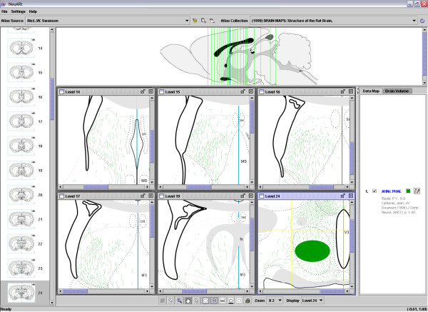
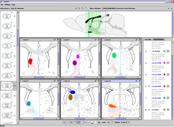
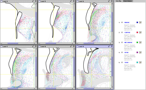
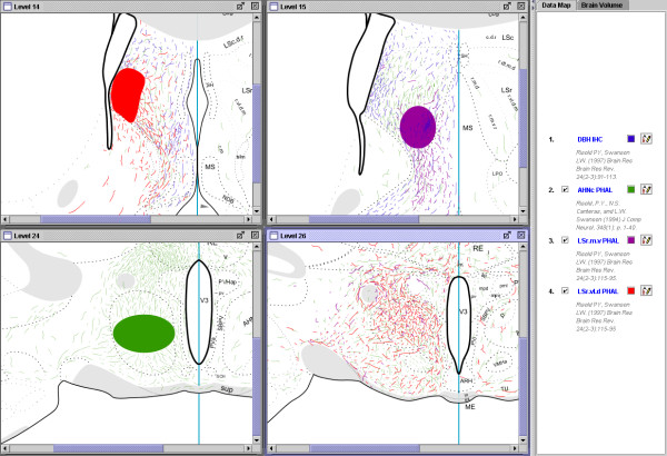
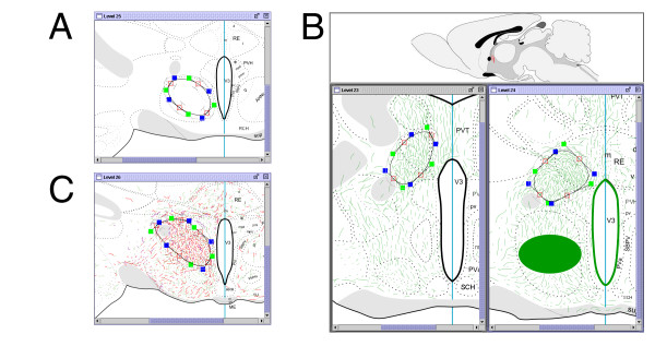
We have developed an atlas-viewing application ('NeuARt II') in the Java language with unique functional properties. These include the ability to use copyrighted atlases as templates within which users may view, save and retrieve data-maps and annotate them with volumetric delineations. NeuARt II also permits users to view multiple levels on multiple atlases at once. Each data-map in this system is simply a stack of vector images with one image per atlas level, so any set of accurate drawings made onto a supported atlas (in vector graphics format) could be uploaded into NeuARt II. Presently the database is populated with a corpus of high-quality neuroanatomical data from the laboratory of Dr Larry Swanson (consisting 64 highly-detailed maps of PHAL tract-tracing experiments, made up of 1039 separate drawings that were published in 27 primary research publications over 17 years). Herein we take selective examples from these data to demonstrate the features of NeuArt II. Our informatics tool permits users to browse, query and compare these maps. The NeuARt II tool operates within a bioinformatics knowledge management platform (called 'NeuroScholar') either as a standalone or a plug-in application.

CONCLUSION

Anatomical localization is fundamental to neuroscientific work and atlases provide an easily-understood framework that is widely used by neuroanatomists and non-neuroanatomists alike. NeuARt II, the neuroinformatics tool presented here, provides an accurate and powerful way of representing neuroanatomical data in the context of commonly-used brain atlases for visualization, comparison and analysis. Furthermore, it provides a framework that supports the delivery and manipulation of mapped data either as a standalone system or as a component in a larger knowledge management system.

摘要

背景

对神经回路的解剖学研究描绘了大脑的基本布线图,产生了对神经科学界具有重大价值的内在空间、高度复杂的数据。已发表的神经解剖学图谱为这些研究提供了一个空间框架。我们基于这些图谱构建了一个信息学框架,用于表示神经解剖学知识。这个框架不仅捕捉了解剖学数据采集和分析的当前方法,还允许在单个系统中对这些研究进行整理、比较和综合。

结果

我们用Java语言开发了一个具有独特功能特性的图谱查看应用程序(“NeuARt II”)。这些特性包括能够将受版权保护的图谱用作模板,用户可以在其中查看、保存和检索数据图,并用体积描绘对其进行注释。NeuARt II还允许用户一次查看多个图谱的多个层面。该系统中的每个数据图只是一堆矢量图像,每个图谱层面有一幅图像,因此任何一组绘制在支持的图谱上(矢量图形格式)的精确图纸都可以上传到NeuARt II中。目前,数据库中填充了来自拉里·斯旺森博士实验室的高质量神经解剖学数据集(由64张PHAL束追踪实验的高度详细地图组成,由1039张单独的图纸组成,这些图纸发表在17年期间的27篇主要研究出版物中)。在此,我们从这些数据中选取了一些示例来展示NeuArt II的特性。我们的信息学工具允许用户浏览、查询和比较这些地图。NeuARt II工具可以作为独立应用程序或插件应用程序在生物信息学知识管理平台(称为“NeuroScholar”)中运行。

结论

解剖定位是神经科学工作的基础,图谱提供了一个易于理解的框架,神经解剖学家和非神经解剖学家都广泛使用。这里介绍的神经信息学工具NeuARt II,提供了一种在常用脑图谱的背景下准确而强大地表示神经解剖学数据的方法,用于可视化、比较和分析。此外,它提供了一个框架,支持将映射数据作为独立系统或更大知识管理系统的组件进行传递和操作。

https://cdn.ncbi.nlm.nih.gov/pmc/blobs/cdce/1770939/08cd0e0c8173/1471-2105-7-531-10.jpg
https://cdn.ncbi.nlm.nih.gov/pmc/blobs/cdce/1770939/4dfc0613f9e4/1471-2105-7-531-1.jpg
https://cdn.ncbi.nlm.nih.gov/pmc/blobs/cdce/1770939/f3391c48f062/1471-2105-7-531-2.jpg
https://cdn.ncbi.nlm.nih.gov/pmc/blobs/cdce/1770939/5b3b3e606cbd/1471-2105-7-531-3.jpg
https://cdn.ncbi.nlm.nih.gov/pmc/blobs/cdce/1770939/13cd610dced2/1471-2105-7-531-4.jpg
https://cdn.ncbi.nlm.nih.gov/pmc/blobs/cdce/1770939/eae2ce00eb97/1471-2105-7-531-5.jpg
https://cdn.ncbi.nlm.nih.gov/pmc/blobs/cdce/1770939/b44b919f091b/1471-2105-7-531-6.jpg
https://cdn.ncbi.nlm.nih.gov/pmc/blobs/cdce/1770939/d661e3cbb56d/1471-2105-7-531-7.jpg
https://cdn.ncbi.nlm.nih.gov/pmc/blobs/cdce/1770939/f6e9ce683587/1471-2105-7-531-8.jpg
https://cdn.ncbi.nlm.nih.gov/pmc/blobs/cdce/1770939/537b46671ca2/1471-2105-7-531-9.jpg
https://cdn.ncbi.nlm.nih.gov/pmc/blobs/cdce/1770939/08cd0e0c8173/1471-2105-7-531-10.jpg

相似文献

1
The NeuARt II system: a viewing tool for neuroanatomical data based on published neuroanatomical atlases.NeuARt II系统:一种基于已发表的神经解剖图谱的神经解剖数据查看工具。
BMC Bioinformatics. 2006 Dec 13;7:531. doi: 10.1186/1471-2105-7-531.
2
The symbolic atlas of the brain: handling non-visual information associated with neuroanatomy.
Med Inform (Lond). 1996 Jan-Mar;21(1):1-21. doi: 10.3109/14639239609009007.
3
Knowledge synthesis with maps of neural connectivity.基于神经连接图谱的知识综合。
Front Neuroinform. 2011 Nov 1;5:24. doi: 10.3389/fninf.2011.00024. eCollection 2011.
4
NeuroTerrain--a client-server system for browsing 3D biomedical image data sets.NeuroTerrain——一个用于浏览三维生物医学图像数据集的客户端-服务器系统。
BMC Bioinformatics. 2007 Feb 5;8:40. doi: 10.1186/1471-2105-8-40.
5
Representation of anatomy in online atlases and databases: a survey and collection of patterns for interface design.在线图谱和数据库中的解剖学呈现:界面设计模式的调查与收集
BMC Dev Biol. 2016 May 21;16(1):18. doi: 10.1186/s12861-016-0116-y.
6
A deformable digital brain atlas system according to Talairach and Tournoux.一种基于塔莱拉赫和图尔诺的可变形数字脑图谱系统。
Med Image Anal. 2004 Mar;8(1):3-22. doi: 10.1016/j.media.2003.06.001.
7
The brain atlas concordance problem: quantitative comparison of anatomical parcellations.脑图谱一致性问题:解剖分区的定量比较。
PLoS One. 2009 Sep 29;4(9):e7200. doi: 10.1371/journal.pone.0007200.
8
The informatics of a C57BL/6J mouse brain atlas.C57BL/6J小鼠脑图谱的信息学
Neuroinformatics. 2003;1(4):397-410. doi: 10.1385/NI:1:4:397.
9
JAtlasView: a Java atlas-viewer for browsing biomedical 3D images and atlases.JAtlasView:一款用于浏览生物医学3D图像和图谱的Java图谱查看器。
BMC Bioinformatics. 2005 Mar 9;6:47. doi: 10.1186/1471-2105-6-47.
10
Constructing and optimizing 3D atlases from 2D data with application to the developing mouse brain.从二维数据构建和优化 3D 图谱及其在发育中老鼠大脑中的应用。
Elife. 2021 Feb 11;10:e61408. doi: 10.7554/eLife.61408.

引用本文的文献

1
Controlling feeding behavior by chemical or gene-directed targeting in the brain: what's so spatial about our methods?通过化学或基因靶向控制大脑中的摄食行为:我们的方法有什么特别的空间性?
Front Neurosci. 2013 Dec 18;7:182. doi: 10.3389/fnins.2013.00182.
2
3D brain atlas reconstructor service--online repository of three-dimensional models of brain structures.3D 脑图谱重构服务——脑结构三维模型在线知识库。
Neuroinformatics. 2013 Oct;11(4):507-18. doi: 10.1007/s12021-013-9199-9.
3
Mapping the connectome: multi-level analysis of brain connectivity.

本文引用的文献

1
Tools for knowledge acquisition within the NeuroScholar system and their application to anatomical tract-tracing data.神经学者系统内的知识获取工具及其在解剖学神经束示踪数据中的应用。
J Biomed Discov Collab. 2006 Aug 8;1:10. doi: 10.1186/1747-5333-1-10.
2
NeuroScholar's electronic laboratory notebook and its application to neuroendocrinology.NeuroScholar电子实验室笔记本及其在神经内分泌学中的应用。
Neuroinformatics. 2006;4(2):139-62. doi: 10.1385/NI:4:2:139.
3
Micro3D: computer program for three-dimensional reconstruction visualization, and analysis of neuronal populations and barin regions.
绘制连接组:大脑连接性的多层次分析。
Front Neuroinform. 2012 May 1;6:14. doi: 10.3389/fninf.2012.00014. eCollection 2012.
4
neuroVIISAS: approaching multiscale simulation of the rat connectome.神经 VIISAS:接近大鼠连接组的多尺度模拟。
Neuroinformatics. 2012 Jul;10(3):243-67. doi: 10.1007/s12021-012-9141-6.
5
Knowledge synthesis with maps of neural connectivity.基于神经连接图谱的知识综合。
Front Neuroinform. 2011 Nov 1;5:24. doi: 10.3389/fninf.2011.00024. eCollection 2011.
6
Comparing histological data from different brains: sources of error and strategies for minimizing them.比较不同大脑的组织学数据:误差来源及将其最小化的策略。
Brain Res Rev. 2009 May;60(2):349-67. doi: 10.1016/j.brainresrev.2009.02.002. Epub 2009 Feb 24.
7
The Brain Connectivity Workshops: moving the frontiers of computational systems neuroscience.大脑连接性研讨会:拓展计算系统神经科学的前沿领域
Neuroimage. 2008 Aug 1;42(1):1-9. doi: 10.1016/j.neuroimage.2008.04.167. Epub 2008 Apr 20.
8
Tools for knowledge acquisition within the NeuroScholar system and their application to anatomical tract-tracing data.神经学者系统内的知识获取工具及其在解剖学神经束示踪数据中的应用。
J Biomed Discov Collab. 2006 Aug 8;1:10. doi: 10.1186/1747-5333-1-10.
Micro3D:用于神经元群体和脑区三维重建、可视化及分析的计算机程序。
Int J Neurosci. 2006 Apr;116(4):515-40. doi: 10.1080/00207450500506025.
4
Three-dimensional chemoarchitecture of the basal forebrain: spatially specific association of cholinergic and calcium binding protein-containing neurons.基底前脑的三维化学结构:胆碱能神经元与含钙结合蛋白神经元的空间特异性关联。
Neuroscience. 2005;136(3):697-713. doi: 10.1016/j.neuroscience.2005.05.019.
5
Projections from bed nuclei of the stria terminalis, anteromedial area: cerebral hemisphere integration of neuroendocrine, autonomic, and behavioral aspects of energy balance.终纹床核的投射,前内侧区:能量平衡的神经内分泌、自主神经和行为方面的大脑半球整合。
J Comp Neurol. 2006 Jan 1;494(1):142-78. doi: 10.1002/cne.20788.
6
Projections from bed nuclei of the stria terminalis, magnocellular nucleus: implications for cerebral hemisphere regulation of micturition, defecation, and penile erection.终纹床核、大细胞核对大脑半球排尿、排便及阴茎勃起调节的投射
J Comp Neurol. 2006 Jan 1;494(1):108-41. doi: 10.1002/cne.20789.
7
Projections from bed nuclei of the stria terminalis, dorsomedial nucleus: implications for cerebral hemisphere integration of neuroendocrine, autonomic, and drinking responses.终纹床核、背内侧核的投射:对神经内分泌、自主神经及饮水反应的大脑半球整合的影响
J Comp Neurol. 2006 Jan 1;494(1):75-107. doi: 10.1002/cne.20790.
8
Projections from the subfornical region of the lateral hypothalamic area.来自下丘脑外侧区穹窿下区域的投射。
J Comp Neurol. 2005 Dec 19;493(3):412-38. doi: 10.1002/cne.20764.
9
Comparison of melanin-concentrating hormone and hypocretin/orexin mRNA expression patterns in a new parceling scheme of the lateral hypothalamic zone.在下丘脑外侧区新分区方案中黑色素浓缩激素与下丘脑泌素/食欲素mRNA表达模式的比较
Neurosci Lett. 2005 Oct 21;387(2):80-4. doi: 10.1016/j.neulet.2005.06.066.
10
Pontine maps linking somatosensory and cerebellar cortices are in register with climbing fiber somatotopy.连接体感皮层和小脑皮层的脑桥图谱与攀缘纤维躯体定位排列一致。
J Neurosci. 2005 Jun 15;25(24):5680-90. doi: 10.1523/JNEUROSCI.0558-05.2005.