• 文献检索
  • 文档翻译
  • 深度研究
  • 学术资讯
  • Suppr Zotero 插件Zotero 插件
  • 邀请有礼
  • 套餐&价格
  • 历史记录
应用&插件
Suppr Zotero 插件Zotero 插件浏览器插件Mac 客户端Windows 客户端微信小程序
定价
高级版会员购买积分包购买API积分包
服务
文献检索文档翻译深度研究API 文档MCP 服务
关于我们
关于 Suppr公司介绍联系我们用户协议隐私条款
关注我们

Suppr 超能文献

核心技术专利:CN118964589B侵权必究
粤ICP备2023148730 号-1Suppr @ 2026

文献检索

告别复杂PubMed语法,用中文像聊天一样搜索,搜遍4000万医学文献。AI智能推荐,让科研检索更轻松。

立即免费搜索

文件翻译

保留排版,准确专业,支持PDF/Word/PPT等文件格式,支持 12+语言互译。

免费翻译文档

深度研究

AI帮你快速写综述,25分钟生成高质量综述,智能提取关键信息,辅助科研写作。

立即免费体验

计算机断层扫描引导下对颅底、咽旁和颞下窝肿块进行细针穿刺抽吸

Computed tomography-directed fine needle aspiration of skull base parapharyngeal and infratemporal fossa masses.

作者信息

Spearman M, Curtin H, Dusenbery D, Janecka I P, Reyna E L

出版信息

Skull Base Surg. 1995;5(4):199-205. doi: 10.1055/s-2008-1058916.

DOI:10.1055/s-2008-1058916
PMID:17170959
原文链接:https://pmc.ncbi.nlm.nih.gov/articles/PMC1656528/
Abstract

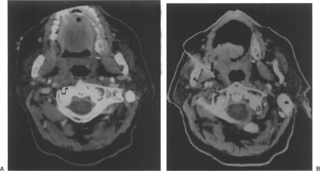
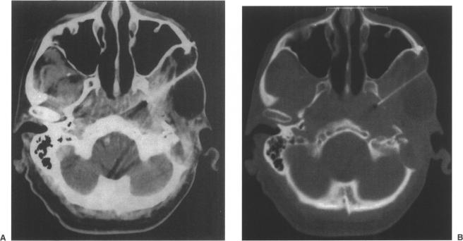
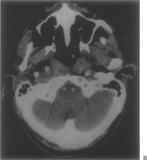
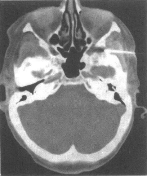
Suspicious findings in the parapharyngeal region on computed tomographic (CT) or magnetic resonance imaging studies can be a diagnostic problem. Blind biopsy through the mucosa can be inadequate, since the abnormality is not directly visible. With CT guidance, fine needle aspiration (FNA) of parapharyngeal masses can be performed with a needle confidently placed within the lesion. Vital structures such as the carotid artery are avoided. We present a series of 33 CT-guided FNA on 30 patients to evaluate the safety and the degree of accuracy of the procedure. Most of the patients had been treated previously for local malignancy. All patients had surgical pathologic study, autopsy, or clinical and imaging follow-up to confirm the FNA cytology results. Twenty of the 33 biopsies were positive for malignant cells, confirming recurrence of the primary head and neck malignancy. Of the 33 CT-directed FNA, 13 were negative for malignant cells. Three of these 13 were found to be false-negative FNA. None of the patients had complications from the procedure. CT directed FNA of masses at the skull base or in the parapharyngeal area can be performed safely. A high degree of accuracy is achieved, with 30 (90.9%) accurate in identifying the presence or absence of malignancy in our series.

摘要

计算机断层扫描(CT)或磁共振成像研究中发现的咽旁区域可疑结果可能是一个诊断难题。由于无法直接看到异常情况,通过黏膜进行盲目活检可能并不充分。在CT引导下,可以用针将其准确置于病变内,对咽旁肿块进行细针穿刺抽吸(FNA)。可避免诸如颈动脉等重要结构。我们对30例患者进行了一系列33次CT引导下的FNA,以评估该操作的安全性和准确性。大多数患者此前曾接受过局部恶性肿瘤治疗。所有患者均接受了手术病理研究、尸检或临床及影像学随访,以确认FNA细胞学结果。33次活检中有20次恶性细胞呈阳性,证实了原发性头颈恶性肿瘤的复发。在33次CT引导下的FNA中,有13次恶性细胞呈阴性。这13次中有3次被发现为FNA假阴性。所有患者均未出现该操作的并发症。可以安全地对颅底或咽旁区域的肿块进行CT引导下的FNA。在我们的系列研究中,其准确性很高,在识别有无恶性肿瘤方面有30次(90.9%)准确。

https://cdn.ncbi.nlm.nih.gov/pmc/blobs/433c/1656528/712b30bdf805/skullbasesurg00026-0017-c.jpg
https://cdn.ncbi.nlm.nih.gov/pmc/blobs/433c/1656528/44c135cd95c6/skullbasesurg00026-0015-a.jpg
https://cdn.ncbi.nlm.nih.gov/pmc/blobs/433c/1656528/adf3c06e07b4/skullbasesurg00026-0015-b.jpg
https://cdn.ncbi.nlm.nih.gov/pmc/blobs/433c/1656528/e92e26d5e787/skullbasesurg00026-0016-a.jpg
https://cdn.ncbi.nlm.nih.gov/pmc/blobs/433c/1656528/43bba21a4816/skullbasesurg00026-0016-b.jpg
https://cdn.ncbi.nlm.nih.gov/pmc/blobs/433c/1656528/b9edf4b38270/skullbasesurg00026-0017-a.jpg
https://cdn.ncbi.nlm.nih.gov/pmc/blobs/433c/1656528/70bbd907e433/skullbasesurg00026-0017-b.jpg
https://cdn.ncbi.nlm.nih.gov/pmc/blobs/433c/1656528/712b30bdf805/skullbasesurg00026-0017-c.jpg

相似文献

1
Computed tomography-directed fine needle aspiration of skull base parapharyngeal and infratemporal fossa masses.计算机断层扫描引导下对颅底、咽旁和颞下窝肿块进行细针穿刺抽吸
Skull Base Surg. 1995;5(4):199-205. doi: 10.1055/s-2008-1058916.
2
Clinical Practice Guideline: Evaluation of the Neck Mass in Adults.临床实践指南:成人颈部肿块的评估
Otolaryngol Head Neck Surg. 2017 Sep;157(2_suppl):S1-S30. doi: 10.1177/0194599817722550.
3
Image-guided fine-needle aspiration of the head and neck: five years' experience.影像引导下的头颈部细针穿刺活检:五年经验
Arch Otolaryngol Head Neck Surg. 1998 Oct;124(10):1155-61. doi: 10.1001/archotol.124.10.1155.
4
Endoscopic ultrasound-guided fine-needle aspiration in 179 cases: the M. D. Anderson Cancer Center experience.179例内镜超声引导下细针穿刺活检:MD安德森癌症中心的经验
Cancer. 2002 Jun 25;96(3):174-80. doi: 10.1002/cncr.10614.
5
Accuracy of fine needle aspiration cytology from breast masses in Thailand.泰国乳腺肿块细针穿刺细胞学检查的准确性。
Asian Pac J Cancer Prev. 2009 Oct-Dec;10(4):623-6.
6
Metabolic PET/CT-guided lung lesion biopsies: impact on diagnostic accuracy and rate of sampling error.代谢PET/CT引导下的肺病变活检:对诊断准确性和采样误差率的影响。
J Nucl Med. 2015 Apr;56(4):518-22. doi: 10.2967/jnumed.113.131466. Epub 2015 Feb 19.
7
Fine-needle aspiration of clinically suspicious palpable breast masses with histopathologic correlation.对临床上可疑的可触及乳腺肿块进行细针穿刺,并进行组织病理学对照。
Am J Surg. 2002 Nov;184(5):410-3. doi: 10.1016/s0002-9610(02)01014-0.
8
Fine needle aspiration cytology in a rural setting.农村地区的细针穿刺细胞学检查。
Acta Cytol. 1998 Jul-Aug;42(4):899-906. doi: 10.1159/000331965.
9
Yield of endoscopic ultrasound-guided fine-needle aspiration of bile duct lesions.内镜超声引导下胆管病变细针穿刺活检的取材率
Endoscopy. 2004 Aug;36(8):715-9. doi: 10.1055/s-2004-825657.
10
Use of fine needle aspiration for solid breast lesions is accurate and cost-effective.对乳腺实性病变使用细针穿刺抽吸术准确且具有成本效益。
Am J Surg. 1997 Dec;174(6):694-6; discussion 697-8. doi: 10.1016/s0002-9610(97)00192-x.

引用本文的文献

1
Tumors of the infratemporal fossa.颞下窝肿瘤。
Skull Base Surg. 2000;10(1):1-9. doi: 10.1055/s-2000-6789.

本文引用的文献

1
Fine needle aspiration cytology of oral and pharyngeal lesions. A study of 45 cases.口腔和咽部病变的细针穿刺细胞学检查:45例研究
Acta Cytol. 1993 May-Jun;37(3):333-42.
2
Peroral fine needle aspiration cytology of parapharyngeal lesions.
Acta Cytol. 1993 Sep-Oct;37(5):694-8.
3
Fine needle aspiration biopsy of intraoral and pharyngeal lesions.
Acta Cytol. 1993 Jul-Aug;37(4):448-50.
4
Biopsy of parapharyngeal space lesions.
咽旁间隙病变活检
Radiology. 1994 Dec;193(3):619-22. doi: 10.1148/radiology.193.3.7972797.
5
Computed tomography-guided aspiration cytologic examination in head and neck lesions.计算机断层扫描引导下的头颈部病变细针穿刺细胞学检查
Arch Otolaryngol. 1984 Sep;110(9):604-7. doi: 10.1001/archotol.1984.00800350046012.
6
Tumors of the parapharyngeal space: preoperative evaluation, diagnosis and surgical approaches.咽旁间隙肿瘤:术前评估、诊断及手术入路
Ann Otol Rhinol Laryngol Suppl. 1981 Jan-Feb;90(1 Pt 4):3-15. doi: 10.1177/00034894810901s301.
7
CT-directed fine needle aspiration biopsies of masses in the head and neck.CT引导下的头颈部肿块细针穿刺活检
Laryngoscope. 1985 Nov;95(11):1382-6. doi: 10.1288/00005537-198511000-00018.
8
Fine needle aspiration cytology in the management ENT of patients.
J Laryngol Otol. 1988 Oct;102(10):909-13. doi: 10.1017/s0022215100106796.
9
Common tumors of the parapharyngeal space: refined imaging diagnosis.咽旁间隙常见肿瘤:精准影像诊断
Radiology. 1988 Oct;169(1):81-5. doi: 10.1148/radiology.169.1.2843942.
10
Fine needle aspiration cytology in the head and neck region.头颈部区域的细针穿刺细胞学检查
Br J Plast Surg. 1989 Jul;42(4):447-51. doi: 10.1016/0007-1226(89)90012-x.