• 文献检索
  • 文档翻译
  • 深度研究
  • 学术资讯
  • Suppr Zotero 插件Zotero 插件
  • 邀请有礼
  • 套餐&价格
  • 历史记录
应用&插件
Suppr Zotero 插件Zotero 插件浏览器插件Mac 客户端Windows 客户端微信小程序
定价
高级版会员购买积分包购买API积分包
服务
文献检索文档翻译深度研究API 文档MCP 服务
关于我们
关于 Suppr公司介绍联系我们用户协议隐私条款
关注我们

Suppr 超能文献

核心技术专利:CN118964589B侵权必究
粤ICP备2023148730 号-1Suppr @ 2026

文献检索

告别复杂PubMed语法,用中文像聊天一样搜索,搜遍4000万医学文献。AI智能推荐,让科研检索更轻松。

立即免费搜索

文件翻译

保留排版,准确专业,支持PDF/Word/PPT等文件格式,支持 12+语言互译。

免费翻译文档

深度研究

AI帮你快速写综述,25分钟生成高质量综述,智能提取关键信息,辅助科研写作。

立即免费体验

时间分辨成像显微镜。磷光和延迟荧光成像。

Time resolved imaging microscopy. Phosphorescence and delayed fluorescence imaging.

作者信息

Marriott G, Clegg R M, Arndt-Jovin D J, Jovin T M

机构信息

Department of Molecular Biology, Max Planck Institute for Biophysical Chemistry, Göttingen, Federal Republic of Germany.

出版信息

Biophys J. 1991 Dec;60(6):1374-87. doi: 10.1016/S0006-3495(91)82175-0.

DOI:10.1016/S0006-3495(91)82175-0
PMID:1723311
原文链接:https://pmc.ncbi.nlm.nih.gov/articles/PMC1260198/
Abstract

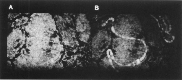
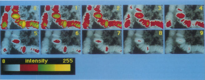
An optical microscope capable of measuring time resolved luminescence (phosphorescence and delayed fluorescence) images has been developed. The technique employs two phase-locked mechanical choppers and a slow-scan scientific CCD camera attached to a normal fluorescence microscope. The sample is illuminated by a periodic train of light pulses and the image is recorded within a defined time interval after the end of each excitation period. The time resolution discriminates completely against light scattering, reflection, autofluorescence, and extraneous prompt fluorescence, which ordinarily decrease contrast in normal fluorescence microscopy measurements. Time resolved image microscopy produces a high contrast image and particular structures can be emphasized by displaying a new parameter, the ratio of the phosphorescence to fluorescence. Objects differing in luminescence decay rates are easily resolved. The lifetime of the long lived luminescence can be measured at each pixel of the microscope image by analyzing a series of images that differ by a variable time delay. The distribution of luminescence decay rates is displayed directly as an image. Several examples demonstrate the utility of the instrument and the complementarity it offers to conventional fluorescence microscopy.

摘要

已开发出一种能够测量时间分辨发光(磷光和延迟荧光)图像的光学显微镜。该技术采用两个锁相机械斩波器和一个连接到普通荧光显微镜的慢扫描科学电荷耦合器件(CCD)相机。样品由周期性的光脉冲序列照射,并在每个激发周期结束后的规定时间间隔内记录图像。时间分辨率完全排除了光散射、反射、自发荧光和外来即时荧光的干扰,这些通常会降低普通荧光显微镜测量中的对比度。时间分辨图像显微镜产生高对比度图像,通过显示一个新参数——磷光与荧光的比率,可以突出特定结构。发光衰减率不同的物体很容易分辨出来。通过分析一系列具有可变时间延迟的图像,可以在显微镜图像的每个像素处测量长寿命发光的寿命。发光衰减率的分布直接显示为一幅图像。几个例子展示了该仪器的实用性及其对传统荧光显微镜的互补性。

https://cdn.ncbi.nlm.nih.gov/pmc/blobs/31b2/1260198/8c082545ad13/biophysj00107-0088-a.jpg
https://cdn.ncbi.nlm.nih.gov/pmc/blobs/31b2/1260198/986bc585eb92/biophysj00107-0084-a.jpg
https://cdn.ncbi.nlm.nih.gov/pmc/blobs/31b2/1260198/d37ba35d4f42/biophysj00107-0085-a.jpg
https://cdn.ncbi.nlm.nih.gov/pmc/blobs/31b2/1260198/88664a368073/biophysj00107-0087-a.jpg
https://cdn.ncbi.nlm.nih.gov/pmc/blobs/31b2/1260198/fe611eb6c963/biophysj00107-0087-b.jpg
https://cdn.ncbi.nlm.nih.gov/pmc/blobs/31b2/1260198/8c082545ad13/biophysj00107-0088-a.jpg

相似文献

1
Time resolved imaging microscopy. Phosphorescence and delayed fluorescence imaging.时间分辨成像显微镜。磷光和延迟荧光成像。
Biophys J. 1991 Dec;60(6):1374-87. doi: 10.1016/S0006-3495(91)82175-0.
2
Time-resolved long-lived luminescence imaging method employing luminescent lanthanide probes with a new microscopy system.采用发光镧系探针和新型显微镜系统的时间分辨长寿命发光成像方法。
J Am Chem Soc. 2007 Nov 7;129(44):13502-9. doi: 10.1021/ja073392j. Epub 2007 Oct 10.
3
High intensity solid-state UV source for time-gated luminescence microscopy.用于时间门控发光显微镜的高强度固态紫外光源。
Cytometry A. 2006 Sep 1;69(9):1020-7. doi: 10.1002/cyto.a.20326.
4
Time-resolved luminescence resonance energy transfer imaging of protein-protein interactions in living cells.活细胞中蛋白质-蛋白质相互作用的时间分辨发光共振能量转移成像
Methods Enzymol. 2012;505:329-45. doi: 10.1016/B978-0-12-388448-0.00025-5.
5
Evaluation of a time-resolved fluorescence microscope using a phosphorescent Pt-porphine model system.使用磷光铂卟啉模型系统对时间分辨荧光显微镜的评估。
Cytometry. 1996 Aug 1;24(4):312-20. doi: 10.1002/(SICI)1097-0320(19960801)24:4<312::AID-CYTO2>3.0.CO;2-H.
6
High resolution detection of fluorescently labeled microorganisms in environmental samples using time-resolved fluorescence microscopy.使用时间分辨荧光显微镜对环境样品中荧光标记的微生物进行高分辨率检测。
FEMS Microbiol Ecol. 2002 Sep 1;41(3):239-45. doi: 10.1111/j.1574-6941.2002.tb00985.x.
7
Fluorescent imaging of pH with optical sensors using time domain dual lifetime referencing.使用时域双寿命参考的光学传感器对pH进行荧光成像。
Anal Chem. 2001 Sep 1;73(17):4354-63. doi: 10.1021/ac0100852.
8
Increasing lanthanide luminescence by use of the RETEL effect.利用RETEL效应增强镧系元素发光。
Cytometry A. 2006 Aug 1;69(8):940-6. doi: 10.1002/cyto.a.20318.
9
Time-resolved delayed luminescence image microscopy using an europium ion chelate complex.使用铕离子螯合物的时间分辨延迟发光图像显微镜技术。
Biophys J. 1994 Sep;67(3):957-65. doi: 10.1016/S0006-3495(94)80597-1.
10
Time-gated luminescence microscopy.时间分辨发光显微镜
Ann N Y Acad Sci. 2008;1130:106-16. doi: 10.1196/annals.1430.032.

引用本文的文献

1
Development of a selective-iodide indicator for live-cell imaging and evaluation of CFTR activity.用于活细胞成像和CFTR活性评估的选择性碘化物指示剂的开发。
Sens Diagn. 2025 Aug 28. doi: 10.1039/d5sd00086f.
2
Development of Time-Resolved Luminescence Measurement Instruments for Biosensing and Bioimaging - An Overview.用于生物传感和生物成像的时间分辨发光测量仪器的发展——综述
Measurement (Lond). 2025 Jun 15;250. doi: 10.1016/j.measurement.2025.117201. Epub 2025 Mar 6.
3
Periodic Light Modulations for Low-Cost Wide-Field Imaging of Luminescence Kinetics Under Ambient Light.

本文引用的文献

1
[TRENDS IN RESEARCH IN THE FIELD OF HISTOLUMINESCENCE].[组织发光领域的研究趋势]
Z Wiss Mikrosk. 1964 Mar;66:164-70.
2
DYNAMICS OF ACRIDINE ORANGE-CELL INTERACTION. II. DYE-INDUCED ULTRASTRUCTURAL CHANGES IN MULTIVESICULAR BODIES (ACRIDINE ORANGE PARTICLES).吖啶橙与细胞相互作用的动力学。II. 染料诱导的多泡体(吖啶橙颗粒)超微结构变化
J Cell Biol. 1964 Apr;21(1):49-62. doi: 10.1083/jcb.21.1.49.
3
DYNAMICS OF ACRIDINE ORANGE-CELL INTERACTION. I. INTERRELATIONSHIPS OF ACRIDINE ORANGE PARTICLES AND CYTOPLASMIC REDDENING.吖啶橙与细胞相互作用的动力学。I. 吖啶橙颗粒与细胞质变红的相互关系。
用于环境光下发光动力学低成本宽视场成像的周期性光调制
Adv Sci (Weinh). 2025 Mar;12(10):e2413291. doi: 10.1002/advs.202413291. Epub 2025 Jan 17.
4
Organic Room Temperature Phosphorescence from BN-Substituted Xanthene Derivatives.硼氮取代呫吨衍生物的有机室温磷光
Angew Chem Int Ed Engl. 2025 Jan 10;64(2):e202414534. doi: 10.1002/anie.202414534. Epub 2024 Nov 11.
5
Accumulated in-situ spectral information analysis of room-temperature phosphorescence with time-gated bioimaging.基于时间分辨生物成像的室温磷光原位光谱信息累积分析
Mater Today Bio. 2024 Sep 14;28:101238. doi: 10.1016/j.mtbio.2024.101238. eCollection 2024 Oct.
6
Low-rate smartphone videoscopy for microsecond luminescence lifetime imaging with machine learning.用于机器学习的微秒级发光寿命成像的低帧率智能手机视频显微镜技术
PNAS Nexus. 2023 Sep 27;2(10):pgad313. doi: 10.1093/pnasnexus/pgad313. eCollection 2023 Oct.
7
Review of Mn-Doped Semiconductor Nanocrystals for Time-Resolved Luminescence Biosensing/Imaging.用于时间分辨发光生物传感/成像的锰掺杂半导体纳米晶体综述。
ACS Appl Nano Mater. 2022 Dec 23;5(12):17413-17435. doi: 10.1021/acsanm.2c04337. Epub 2022 Nov 30.
8
AIE-active polyanetholesulfonic acid sodium salts with room-temperature phosphorescence characteristics for Fe detection.具有室温磷光特性的用于检测铁的聚集诱导发光活性聚苯乙烯磺酸钠盐。
RSC Adv. 2018 Sep 4;8(54):31231-31236. doi: 10.1039/c8ra06178e. eCollection 2018 Aug 30.
9
Optical Properties of Mn-Doped CuGa(In)S-ZnS Nanocrystals (NCs): Effects of Host NC and Mn Concentration.锰掺杂的CuGa(In)S-ZnS纳米晶体(NCs)的光学性质:主体纳米晶体和锰浓度的影响
Nanomaterials (Basel). 2022 Mar 17;12(6):994. doi: 10.3390/nano12060994.
10
Mn-Doped AgZnInS/ZnS Nanocrystals (NCs): Effects of Zn Etching on the NC Optical Properties.锰掺杂的AgZnInS/ZnS纳米晶体(NCs):锌蚀刻对NC光学性质的影响
Opt Mater (Amst). 2022 Jan;123. doi: 10.1016/j.optmat.2021.111941. Epub 2021 Dec 28.
J Cell Biol. 1963 Aug;18(2):237-50. doi: 10.1083/jcb.18.2.237.
4
Europium as a label in time-resolved immunofluorometric assays.铕作为时间分辨免疫荧光分析中的标记物。
Anal Biochem. 1984 Mar;137(2):335-43. doi: 10.1016/0003-2697(84)90095-2.
5
Depolarization of fluorescence depletion. A microscopic method for measuring rotational diffusion of membrane proteins on the surface of a single cell.荧光耗尽去极化。一种用于测量单个细胞表面膜蛋白旋转扩散的显微方法。
FEBS Lett. 1981 Sep 28;132(2):252-6. doi: 10.1016/0014-5793(81)81172-6.
6
[An installation for the study of phosphorescence in micro-objects].[用于研究微观物体中磷光现象的装置]
Tsitologiia. 1966 Sep-Oct;8(5):677-81.
7
The low temperature luminescence of complexes of acridine orange with polyelectrolytes.吖啶橙与聚电解质复合物的低温发光
Biochim Biophys Acta. 1970;209(2):306-19. doi: 10.1016/0005-2787(70)90729-x.
8
Fluorescence microscopy of lysosomes and related structures in living cells.活细胞中溶酶体及相关结构的荧光显微镜检查。
Nature. 1966 Oct 22;212(5060):425-6. doi: 10.1038/212425a0.
9
Fluorescence digital imaging microscopy in cell biology.细胞生物学中的荧光数字成像显微镜技术。
Science. 1985 Oct 18;230(4723):247-56. doi: 10.1126/science.4048934.
10
Proflavin binding to poly[d(A-T)] and poly[d(A-br5U)]: triplet state and temperature-jump kinetics.原黄素与聚[d(A-T)]和聚[d(A-br5U)]的结合:三重态和温度跃变动力学
Biochemistry. 1986 Jul 15;25(14):3995-4007. doi: 10.1021/bi00362a004.