Spahn Gunter, Plettenberg Holger, Kahl Enrico, Klinger Hans M, Mückley Thomas, Hofmann Gunther O
Center of Traumatology and Orthopaedic Surgery, Eisenach, Germany.
BMC Musculoskelet Disord. 2007 May 29;8:47. doi: 10.1186/1471-2474-8-47.
Arthroscopy is a highly sensitive method of evaluating high-grade cartilage lesions but the detection of low-grade lesions is often is unreliable. Objective measurements are required. A novel NIRS (near-infrared-spectroscopy) device for detection of low-grade cartilage defects was evaluated in a preliminary clinical study.
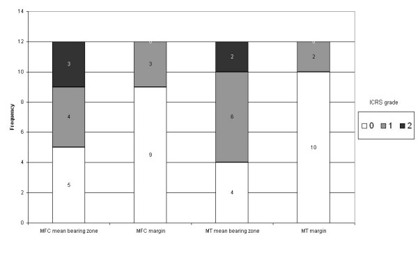
In 12 patients who had undergone arthroscopy, the cartilage lesions within the medial knee compartment were classified according to the ICRS protocol. With a NIR spectrometer system and an optical probe, similar in design to a hook used for routine arthroscopy, the optical properties of cartilage were measured during arthroscopy.
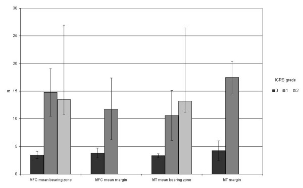
The mean ratio of 2 NIR absorption bands of intact cartilage 3.8 (range 2.3 to 8.7).was significantly lower than that of cartilage with grade 1 lesions (12.8, range 4.8 to 19.6) and grade 2 lesions (13.4, range 10.4 to 15.4).No differences were observed between grade 1 and grade 2 lesions.
NIRS can be used to distinguish between ICRS grade 1 lesions and healthy cartilage during arthroscopic surgeries. The results of this clinical study demonstrate the potential of NIRS to objectify classical arthroscopic grading systems.
关节镜检查是评估高级别软骨损伤的一种高度敏感的方法,但对低级别损伤的检测往往不可靠。需要客观测量。在一项初步临床研究中对一种用于检测低级别软骨缺损的新型近红外光谱(NIRS)设备进行了评估。
在12例接受关节镜检查的患者中,根据国际软骨修复协会(ICRS)方案对膝关节内侧间室的软骨损伤进行分类。使用一个近红外光谱仪系统和一个光学探头(其设计类似于用于常规关节镜检查的钩子),在关节镜检查期间测量软骨的光学特性。
完整软骨的2个近红外吸收带的平均比值为3.8(范围为2.3至8.7),显著低于1级损伤软骨(12.8,范围为4.8至19.6)和2级损伤软骨(13.4,范围为10.4至15.4)。1级和2级损伤之间未观察到差异。
在关节镜手术期间,近红外光谱可用于区分ICRS 1级损伤和健康软骨。这项临床研究的结果证明了近红外光谱使经典关节镜分级系统客观化的潜力。