Kandel Shital, Querido William, Falcon Jessica M, Zlotnick Hannah M, Locke Ryan C, Stoeckl Brendan, Patel Jay M, Patil Chetan A, Mauck Robert L, Pleshko Nancy
Department of Bioengineering, Temple University, Philadelphia, PA, United States.
Department of Bioengineering, University of Pennsylvania, Philadelphia, PA, United States.
Front Bioeng Biotechnol. 2022 Aug 23;10:885369. doi: 10.3389/fbioe.2022.885369. eCollection 2022.
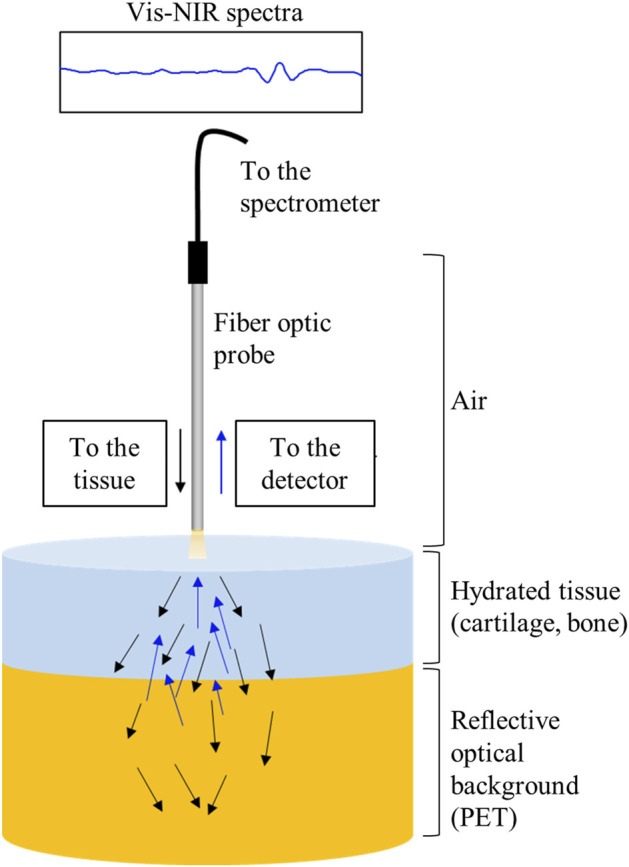
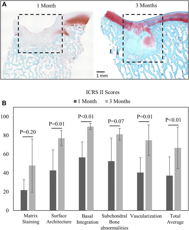
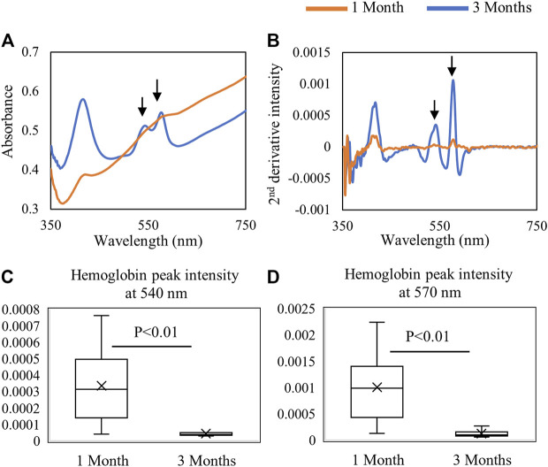
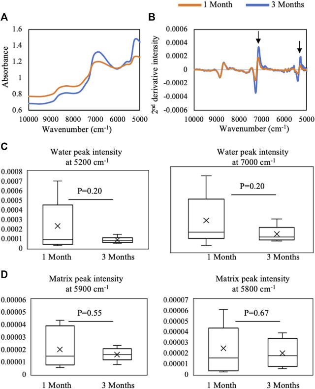
Standard assessment of cartilage repair progression by visual arthroscopy can be subjective and may result in suboptimal evaluation. Visible-near infrared (Vis-NIR) fiber optic spectroscopy of joint tissues, including articular cartilage and subchondral bone, provides an objective approach for quantitative assessment of tissue composition. Here, we applied this technique in the 350-2,500 nm spectral region to identify spectral markers of osteochondral tissue during repair with the overarching goal of developing a new approach to monitor repair of cartilage defects . Full thickness chondral defects were created in Yucatan minipigs using a 5-mm biopsy punch, and microfracture (MFx) was performed as a standard technique to facilitate repair. Tissues were evaluated at 1 month (in adult pigs) and 3 months (in juvenile pigs) post-surgery by spectroscopy and histology. After euthanasia, Vis-NIR spectra were collected from the defect region. Additional spectroscopy experiments were carried out to aid in spectral interpretation. Osteochondral tissues were dissected from the joint and evaluated using the conventional International Cartilage Repair Society (ICRS) II histological scoring system, which showed lower scores for the 1-month than the 3-month repair tissues. In the visible spectral region, hemoglobin absorbances at 540 and 570 nm were significantly higher in spectra from 1-month repair tissue than 3-month repair tissue, indicating a reduction of blood in the more mature repair tissue. In the NIR region, we observed qualitative differences between the two groups in spectra taken from the defect, but differences did not reach significance. Furthermore, spectral data also indicated that the hydrated environment of the joint tissue may interfere with evaluation of tissue water absorbances in the NIR region. Together, these data provide support for further investigation of the visible spectral region for assessment of longitudinal repair of cartilage defects, which would enable assessment during routine arthroscopy, particularly in a hydrated environment.
通过视觉关节镜对软骨修复进展进行的标准评估可能具有主观性,并且可能导致评估效果不佳。对包括关节软骨和软骨下骨在内的关节组织进行可见-近红外(Vis-NIR)光纤光谱分析,为组织成分的定量评估提供了一种客观方法。在此,我们将该技术应用于350-2500nm光谱区域,以识别修复过程中骨软骨组织的光谱标记物,总体目标是开发一种监测软骨缺损修复的新方法。使用5mm活检打孔器在尤卡坦小型猪中制造全层软骨缺损,并采用微骨折(MFx)作为促进修复的标准技术。在术后1个月(成年猪)和3个月(幼年猪)通过光谱学和组织学对组织进行评估。安乐死后,从缺损区域收集Vis-NIR光谱。进行了额外的光谱学实验以辅助光谱解释。从关节中解剖出骨软骨组织,并使用传统的国际软骨修复协会(ICRS)II组织学评分系统进行评估,结果显示1个月修复组织的评分低于3个月修复组织。在可见光谱区域,1个月修复组织光谱中540和570nm处的血红蛋白吸光度显著高于3个月修复组织,表明在更成熟的修复组织中血液减少。在近红外区域,我们观察到两组从缺损处采集的光谱存在定性差异,但差异未达到显著水平。此外,光谱数据还表明关节组织的水合环境可能会干扰近红外区域组织吸水度的评估。总之,这些数据为进一步研究可见光谱区域以评估软骨缺损的纵向修复提供了支持,这将能够在常规关节镜检查期间进行评估,特别是在水合环境中。