Al Absi Mohammad, Qais Abdul Malik, Al Katta Mohammad, Gafour Mohammed, Al-Wadan Ali Hamoud
Radiology Department, Sana'a University, Sana'a, Yemen.
Ann Saudi Med. 2007 May-Jun;27(3):161-5. doi: 10.5144/0256-4947.2007.161.
Conventional methods of radiographic examination are often unsatisfactory for identifying worms in the biliary tract. Ultrasonography is a non-invasive, quick and safe procedure known to have diagnostic accuracy. We studied the ultrasonographic appearances of biliary ascariasis and the role of ultrasonography in diagnosis and management.
In a prospective 5-year study, a sonographic diagnosis of biliary ascariasis was made on 46 Yemeni patients. The diagnosis was based mainly on sonographic appearances supported by clinical and laboratory results and proved by outcome of either surgical or medical management or spontaneous exit of worms. Follow-up ultrasound was performed for all patients to confirm the diagnosis and to monitor management.
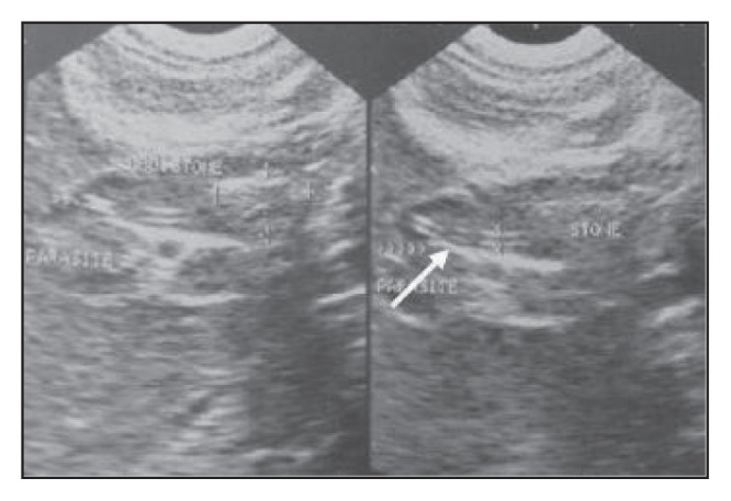
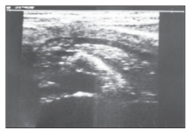
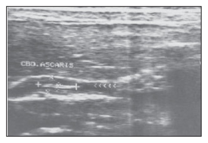
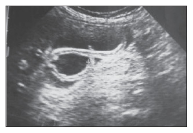
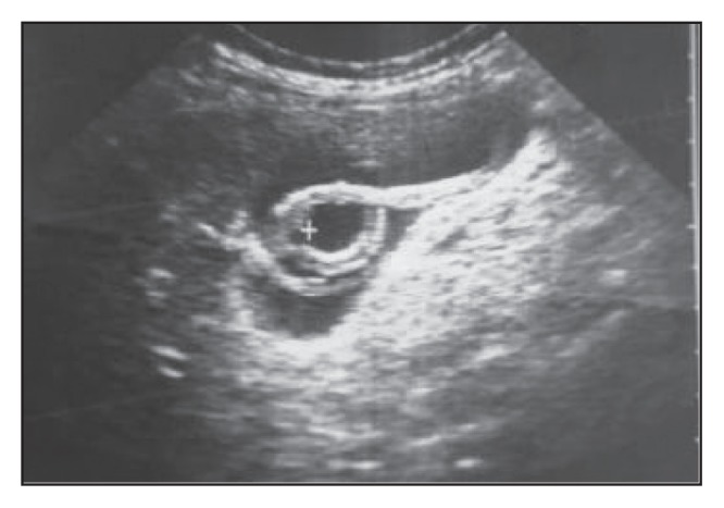
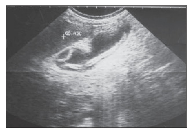
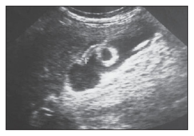
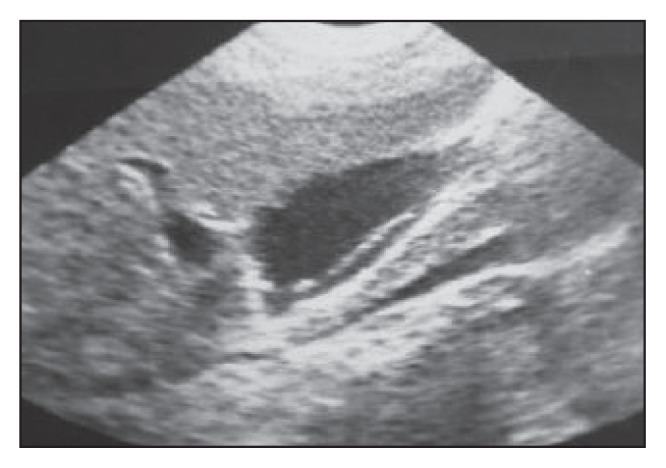
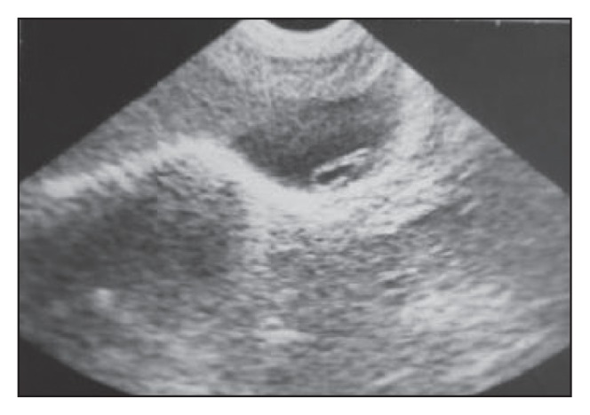
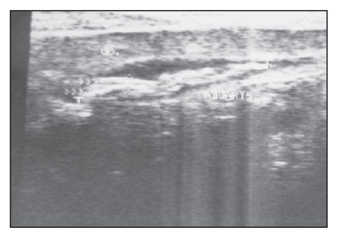
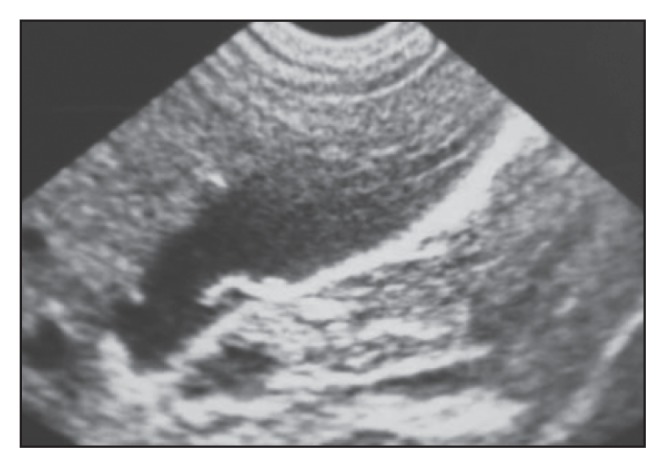
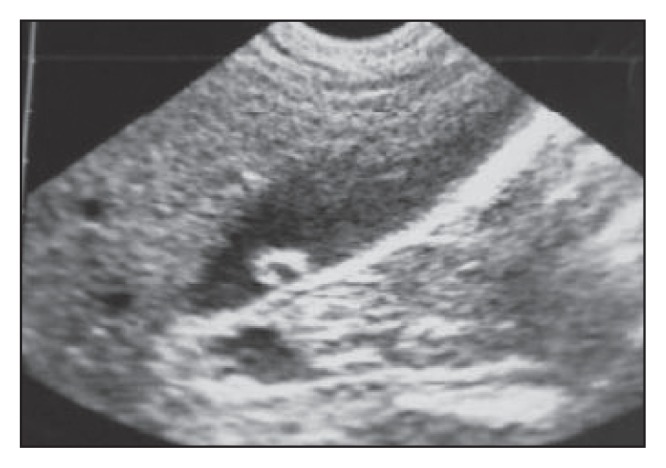
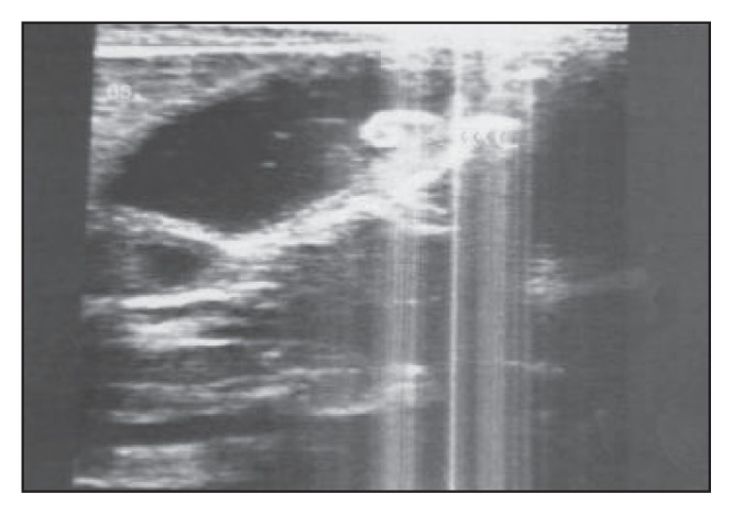
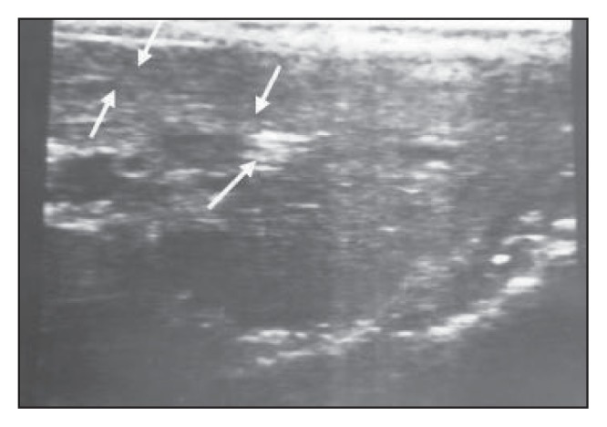
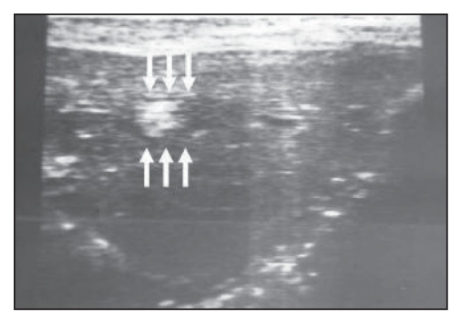
Parasites were present in the dilated main bile duct in 23 patients, in the gallbladder in 12 patients, in the intrahepatic ducts in 6 patients, in the main pancreatic duct in 4 patients and as an intrahepatic abscess in one patient. The characteristic appearance of Ascaris lumbricoides was as single or multiple echogenic non- shadowing linear or curved strips with or without echoic tubular central lines that represent the digestive tracts of the worm. A spaghetti-like appearance was seen in 9 patients and amorphous fragments were seen in 2 patients. Sixteen patients underwent surgery, 20 patients were treated medically (including spontaneous exit of the worm in 7 patients without treatment) and in 10 patients worms were extracted by endoscopic retrograde cholangiopancreatography.
Follow-up ultrasound was found to be effective in confirming the diagnosis and monitoring management.
传统的放射学检查方法在识别胆道蛔虫方面常常不尽人意。超声检查是一种无创、快速且安全的检查方法,已知具有诊断准确性。我们研究了胆道蛔虫病的超声表现以及超声检查在诊断和治疗中的作用。
在一项为期5年的前瞻性研究中,对46例也门患者进行了超声诊断胆道蛔虫病。诊断主要基于超声表现,并得到临床和实验室结果的支持,且通过手术或药物治疗结果或蛔虫自行排出得到证实。对所有患者进行了随访超声检查,以确认诊断并监测治疗情况。
23例患者的扩张胆总管内有寄生虫,12例患者的胆囊内有寄生虫,6例患者的肝内胆管内有寄生虫,4例患者的主胰管内有寄生虫,1例患者的肝内出现脓肿。蛔虫的特征性表现为单条或多条强回声、无阴影的线性或弯曲条带,有或无代表蛔虫消化道的回声管状中心线。9例患者出现类似意大利面条的表现,2例患者出现无定形碎片。16例患者接受了手术,20例患者接受了药物治疗(包括7例未经治疗蛔虫自行排出的患者),10例患者通过内镜逆行胰胆管造影术取出蛔虫。
随访超声检查在确认诊断和监测治疗方面被发现是有效的。