• 文献检索
  • 文档翻译
  • 深度研究
  • 学术资讯
  • Suppr Zotero 插件Zotero 插件
  • 邀请有礼
  • 套餐&价格
  • 历史记录
应用&插件
Suppr Zotero 插件Zotero 插件浏览器插件Mac 客户端Windows 客户端微信小程序
定价
高级版会员购买积分包购买API积分包
服务
文献检索文档翻译深度研究API 文档MCP 服务
关于我们
关于 Suppr公司介绍联系我们用户协议隐私条款
关注我们

Suppr 超能文献

核心技术专利:CN118964589B侵权必究
粤ICP备2023148730 号-1Suppr @ 2026

文献检索

告别复杂PubMed语法,用中文像聊天一样搜索,搜遍4000万医学文献。AI智能推荐,让科研检索更轻松。

立即免费搜索

文件翻译

保留排版,准确专业,支持PDF/Word/PPT等文件格式,支持 12+语言互译。

免费翻译文档

深度研究

AI帮你快速写综述,25分钟生成高质量综述,智能提取关键信息,辅助科研写作。

立即免费体验

使用特征线并行化和归一化互信息的植入后计算机断层扫描-磁共振前列腺图像配准

Post-implant computed tomography-magnetic resonance prostate image registration using feature line parallelization and normalized mutual information.

作者信息

Vidakovic Sandra, Jans Hans S, Alexander Abe, Sloboda Ron S

机构信息

Cross Cancer Institute, Department of Medical Physics, Edmonton, Alberta, Canada.

出版信息

J Appl Clin Med Phys. 2006 Jul 5;8(1):21-32. doi: 10.1120/jacmp.v8i1.2351.

DOI:10.1120/jacmp.v8i1.2351
PMID:17592452
原文链接:https://pmc.ncbi.nlm.nih.gov/articles/PMC5722399/
Abstract

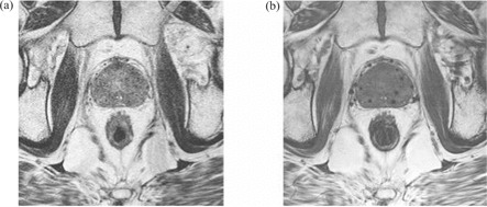
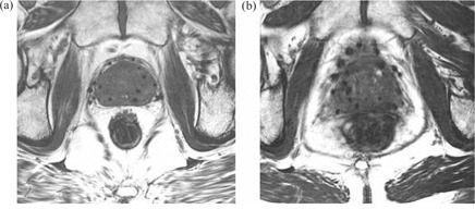
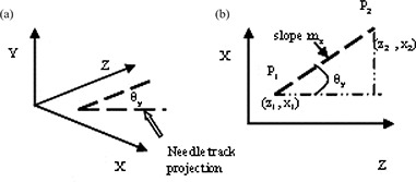
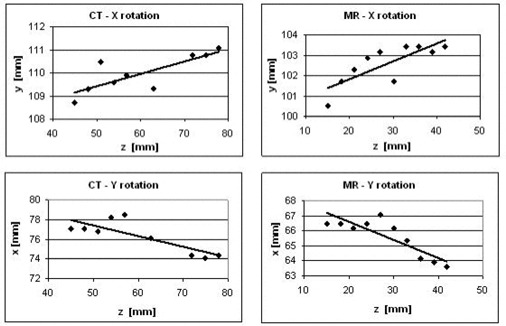
Post-implant dosimetry for permanent prostate brachytherapy is typically performed using computed tomography (CT) images, for which the clear visualization of soft tissue structures is problematic. Registration of CT and magnetic resonance (MR) image volumes can improve the definition of all structures of interest (soft tissues, bones, and seeds) in the joint image set. In the present paper, we describe a novel two-stage rigid-body registration algorithm that consists of (1) parallelization of straight lines fit to image features running primarily in the superior-inferior (Z) direction, followed by (2) normalized mutual information registration. The first stage serves to fix rotation angles about the anterior-posterior (Y) and left-right (X) directions, and the second stage determines the remaining Z-axis rotation angle and the X, Y, Z translation values. The new algorithm was applied to CT and 1.5T MR (T2-weighted and balanced fast-field echo sequences) axial image sets for three patients acquired four weeks after prostate brachytherapy using 125I seeds. Image features used for the stage 1 parallelization were seed trains in CT and needle tracks and seed voids in MR. Simulated datasets were also created to further investigate algorithm performance. Clinical image volumes were successfully registered using the two-stage approach to within a root-mean-squares (RMS) distance of <1.5 mm, provided that some pubic bone and anterior rectum were included in the registration volume of interest and that no motion artifact was apparent. This level of accuracy is comparable to that obtained for the same clinical datasets using the Procrustes algorithm. Unlike Procrustes, the new algorithm can be almost fully automated, and hence we conclude that its further development for application in post-implant dosimetry is warranted.

摘要

永久性前列腺近距离放射治疗植入后的剂量测定通常使用计算机断层扫描(CT)图像进行,而在这些图像中软组织结构的清晰可视化存在问题。CT图像体积与磁共振(MR)图像体积的配准可以改善联合图像集中所有感兴趣结构(软组织、骨骼和籽源)的清晰度。在本文中,我们描述了一种新颖的两阶段刚体配准算法,该算法包括:(1)对主要沿上下(Z)方向运行的图像特征拟合直线进行并行化处理,随后进行(2)归一化互信息配准。第一阶段用于确定围绕前后(Y)和左右(X)方向的旋转角度,第二阶段确定剩余的Z轴旋转角度以及X、Y、Z平移值。该新算法应用于三位患者在前列腺近距离放射治疗使用¹²⁵I籽源四周后获取的CT和1.5T MR(T2加权和平衡快速场回波序列)轴向图像集。用于第一阶段并行化处理的图像特征在CT中为籽源链,在MR中为针道和籽源空洞。还创建了模拟数据集以进一步研究算法性能。使用两阶段方法成功地将临床图像体积配准到均方根(RMS)距离<1.5毫米以内,前提是感兴趣的配准体积中包含一些耻骨和直肠前部且无明显运动伪影。这种精度水平与使用普罗克汝斯算法对相同临床数据集所获得的精度相当。与普罗克汝斯算法不同,新算法几乎可以完全自动化,因此我们得出结论,有必要对其进行进一步开发以应用于植入后剂量测定。

https://cdn.ncbi.nlm.nih.gov/pmc/blobs/b111/5722399/e21831bace01/ACM2-8-021-g005.jpg
https://cdn.ncbi.nlm.nih.gov/pmc/blobs/b111/5722399/971b9e2ba731/ACM2-8-021-g001.jpg
https://cdn.ncbi.nlm.nih.gov/pmc/blobs/b111/5722399/3049cd5c2c47/ACM2-8-021-g002.jpg
https://cdn.ncbi.nlm.nih.gov/pmc/blobs/b111/5722399/8c9fd6e86f94/ACM2-8-021-g003.jpg
https://cdn.ncbi.nlm.nih.gov/pmc/blobs/b111/5722399/f137db65e4d4/ACM2-8-021-g004.jpg
https://cdn.ncbi.nlm.nih.gov/pmc/blobs/b111/5722399/e21831bace01/ACM2-8-021-g005.jpg

相似文献

1
Post-implant computed tomography-magnetic resonance prostate image registration using feature line parallelization and normalized mutual information.使用特征线并行化和归一化互信息的植入后计算机断层扫描-磁共振前列腺图像配准
J Appl Clin Med Phys. 2006 Jul 5;8(1):21-32. doi: 10.1120/jacmp.v8i1.2351.
2
Use and uncertainties of mutual information for computed tomography/ magnetic resonance (CT/MR) registration post permanent implant of the prostate.前列腺永久性植入术后计算机断层扫描/磁共振成像(CT/MR)配准中互信息的应用与不确定性
Med Phys. 2005 Feb;32(2):473-82. doi: 10.1118/1.1851920.
3
Towards integrating functional imaging in the treatment of prostate cancer with radiation: the registration of the MR spectroscopy imaging to ultrasound/CT images and its implementation in treatment planning.迈向将功能成像整合于前列腺癌放射治疗中:磁共振波谱成像与超声/CT图像的配准及其在治疗计划中的应用
Int J Radiat Oncol Biol Phys. 2002 Dec 1;54(5):1558-64. doi: 10.1016/s0360-3016(02)03805-1.
4
The use of mutual information in registration of CT and MRI datasets post permanent implant.互信息在永久性植入后CT和MRI数据集配准中的应用。
Brachytherapy. 2004;3(2):61-70. doi: 10.1016/j.brachy.2004.06.001.
5
Narrow band deformable registration of prostate magnetic resonance imaging, magnetic resonance spectroscopic imaging, and computed tomography studies.前列腺磁共振成像、磁共振波谱成像和计算机断层扫描研究的窄带可变形配准
Int J Radiat Oncol Biol Phys. 2005 Jun 1;62(2):595-605. doi: 10.1016/j.ijrobp.2005.02.001.
6
Prostate seed implant quality assessment using MR and CT image fusion.
Int J Radiat Oncol Biol Phys. 1999 Jan 1;43(1):67-72. doi: 10.1016/s0360-3016(98)00372-1.
7
A software system for interventional magnetic resonance image-guided prostate brachytherapy.一种用于介入性磁共振图像引导前列腺近距离放射治疗的软件系统。
Comput Aided Surg. 2000;5(6):401-13. doi: 10.1002/igs.1002.
8
Inter-slice bidirectional registration-based segmentation of the prostate gland in MR and CT image sequences.基于层间双向配准的 MR 和 CT 图像序列中前列腺分割。
Med Phys. 2013 Dec;40(12):123503. doi: 10.1118/1.4829511.
9
Comparison of CT and MR-CT fusion for prostate post-implant dosimetry.CT 与 MR-CT 融合在前列腺植入后剂量学中的比较。
Int J Radiat Oncol Biol Phys. 2012 Apr 1;82(5):1912-7. doi: 10.1016/j.ijrobp.2011.01.064. Epub 2011 May 6.
10
Prostate post-implant dosimetry: interobserver variability in seed localisation, contouring and fusion.前列腺植入后剂量学:种子定位、勾画和融合的观察者间变异性。
Radiother Oncol. 2012 Aug;104(2):192-8. doi: 10.1016/j.radonc.2012.06.014. Epub 2012 Jul 31.

引用本文的文献

1
Investigation of the clinical inter-observer bias in prostate fiducial marker image registration between CT and MR images.探讨 CT 和 MR 图像中前列腺基准标记物图像配准的临床观察者间偏倚。
Radiat Oncol. 2021 Aug 16;16(1):150. doi: 10.1186/s13014-021-01865-8.
2
CT AND MRI FUSION FOR POSTIMPLANT PROSTATE BRACHYTHERAPY EVALUATION.用于植入后前列腺近距离放射治疗评估的CT与MRI融合技术
Proc IEEE Int Symp Biomed Imaging. 2016 Apr;2016:625-628. doi: 10.1109/ISBI.2016.7493345. Epub 2016 Jun 16.
3
Comparison of manual and automatic MR-CT registration for radiotherapy of prostate cancer.

本文引用的文献

1
Sequential evaluation of prostate edema after permanent seed prostate brachytherapy using CT-MRI fusion.使用CT-MRI融合技术对永久性粒子植入前列腺近距离放疗后前列腺水肿进行序贯评估。
Int J Radiat Oncol Biol Phys. 2005 Jul 15;62(4):974-80. doi: 10.1016/j.ijrobp.2004.12.012.
2
Use and uncertainties of mutual information for computed tomography/ magnetic resonance (CT/MR) registration post permanent implant of the prostate.前列腺永久性植入术后计算机断层扫描/磁共振成像(CT/MR)配准中互信息的应用与不确定性
Med Phys. 2005 Feb;32(2):473-82. doi: 10.1118/1.1851920.
3
The use of mutual information in registration of CT and MRI datasets post permanent implant.
手动和自动磁共振-计算机断层扫描配准在前列腺癌放疗中的比较。
J Appl Clin Med Phys. 2016 May 8;17(3):294-303. doi: 10.1120/jacmp.v17i3.6088.
4
Effect of a urinary catheter on seed position and rectal and bladder doses in CT-based post-implant dosimetry for prostate cancer brachytherapy.导尿管对前列腺癌近距离放疗基于CT的植入后剂量测定中籽源位置以及直肠和膀胱剂量的影响。
J Contemp Brachytherapy. 2015 Jun;7(3):211-7. doi: 10.5114/jcb.2015.52624. Epub 2015 Jun 29.
5
A new two-step accurate CT-MRI fusion technique for post-implant prostate cancer.一种用于植入后前列腺癌的新型两步精确CT-MRI融合技术。
J Contemp Brachytherapy. 2015 Apr;7(2):117-21. doi: 10.5114/jcb.2015.51290. Epub 2015 Apr 30.
6
Feasibility of vibro-acoustography with a quasi-2D ultrasound array transducer for detection and localizing of permanent prostate brachytherapy seeds: a pilot ex vivo study.使用准二维超声阵列换能器进行振动声学造影以检测和定位永久性前列腺近距离治疗种子源的可行性:一项离体初步研究。
Med Phys. 2014 Sep;41(9):092902. doi: 10.1118/1.4893532.
7
Elastic registration of multimodal prostate MRI and histology via multiattribute combined mutual information.基于多属性联合互信息的多模态前列腺 MRI 和组织病理学的弹性配准。
Med Phys. 2011 Apr;38(4):2005-18. doi: 10.1118/1.3560879.
8
Systematisation of spatial uncertainties for comparison between a MR and a CT-based radiotherapy workflow for prostate treatments.对基于 MR 和 CT 的前列腺治疗放射治疗工作流程之间的空间不确定性进行系统分析。
Radiat Oncol. 2009 Nov 17;4:54. doi: 10.1186/1748-717X-4-54.
互信息在永久性植入后CT和MRI数据集配准中的应用。
Brachytherapy. 2004;3(2):61-70. doi: 10.1016/j.brachy.2004.06.001.
4
A comparison of CT scan to transrectal ultrasound-measured prostate volume in untreated prostate cancer.未经治疗的前列腺癌中CT扫描与经直肠超声测量前列腺体积的比较。
Int J Radiat Oncol Biol Phys. 2003 Sep 1;57(1):29-32. doi: 10.1016/s0360-3016(03)00509-1.
5
A systematic study of imaging uncertainties and their impact on 125I prostate brachytherapy dose evaluation.
Med Phys. 2003 Jul;30(7):1897-908. doi: 10.1118/1.1586451.
6
The effect of interobserver differences in post-implant prostate CT image interpretation on dosimetric parameters.
Med Phys. 2003 Jun;30(6):1096-102. doi: 10.1118/1.1576232.
7
Magnetic resonance imaging in the radiation treatment planning of localized prostate cancer using intra-prostatic fiducial markers for computed tomography co-registration.使用前列腺内基准标记进行计算机断层扫描共配准的局部前列腺癌放射治疗计划中的磁共振成像。
Radiother Oncol. 2003 Feb;66(2):217-24. doi: 10.1016/s0167-8140(02)00407-3.
8
[CT and MR image fusion using two different methods after prostate brachytherapy: impact on post-implant dosimetric assessment].[前列腺近距离放疗后使用两种不同方法进行CT与MR图像融合:对植入后剂量学评估的影响]
Cancer Radiother. 2003 Feb;7(1):9-16. doi: 10.1016/s1278-3218(02)00285-8.
9
Comparison of MRI pulse sequences in defining prostate volume after permanent implantation.永久性植入后MRI脉冲序列在确定前列腺体积方面的比较。
Int J Radiat Oncol Biol Phys. 2002 Nov 1;54(3):703-11. doi: 10.1016/s0360-3016(02)02991-7.
10
Prostate seed implant quality assessment using MR and CT image fusion.
Int J Radiat Oncol Biol Phys. 1999 Jan 1;43(1):67-72. doi: 10.1016/s0360-3016(98)00372-1.