• 文献检索
  • 文档翻译
  • 深度研究
  • 学术资讯
  • Suppr Zotero 插件Zotero 插件
  • 邀请有礼
  • 套餐&价格
  • 历史记录
应用&插件
Suppr Zotero 插件Zotero 插件浏览器插件Mac 客户端Windows 客户端微信小程序
定价
高级版会员购买积分包购买API积分包
服务
文献检索文档翻译深度研究API 文档MCP 服务
关于我们
关于 Suppr公司介绍联系我们用户协议隐私条款
关注我们

Suppr 超能文献

核心技术专利:CN118964589B侵权必究
粤ICP备2023148730 号-1Suppr @ 2026

文献检索

告别复杂PubMed语法,用中文像聊天一样搜索,搜遍4000万医学文献。AI智能推荐,让科研检索更轻松。

立即免费搜索

文件翻译

保留排版,准确专业,支持PDF/Word/PPT等文件格式,支持 12+语言互译。

免费翻译文档

深度研究

AI帮你快速写综述,25分钟生成高质量综述,智能提取关键信息,辅助科研写作。

立即免费体验

手动和自动磁共振-计算机断层扫描配准在前列腺癌放疗中的比较。

Comparison of manual and automatic MR-CT registration for radiotherapy of prostate cancer.

机构信息

Aalborg University.

出版信息

J Appl Clin Med Phys. 2016 May 8;17(3):294-303. doi: 10.1120/jacmp.v17i3.6088.

DOI:10.1120/jacmp.v17i3.6088
PMID:27167285
原文链接:https://pmc.ncbi.nlm.nih.gov/articles/PMC5690943/
Abstract

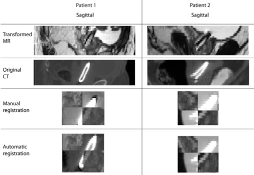
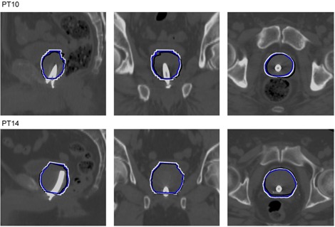
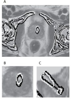
In image-guided radiotherapy (IGRT) of prostate cancer, delineation of the clini-cal target volume (CTV) often relies on magnetic resonance (MR) because of its good soft-tissue visualization. Registration of MR and computed tomography (CT) is required in order to add this accurate delineation to the dose planning CT. An automatic approach for local MR-CT registration of the prostate has previously been developed using a voxel property-based registration as an alternative to a manual landmark-based registration. The aim of this study is to compare the two registration approaches and to investigate the clinical potential for replacing the manual registration with the automatic registration. Registrations and analysis were performed for 30 prostate cancer patients treated with IGRT using a Ni-Ti prostate stent as a fiducial marker. The comparison included computing translational and rotational differences between the approaches, visual inspection, and computing the overlap of the CTV. The computed mean translational difference was 1.65, 1.60, and 1.80mm and the computed mean rotational difference was 1.51°, 3.93°, and 2.09° in the superior/inferior, anterior/posterior, and medial/lateral direction, respectively. The sensitivity of overlap was 87%. The results demonstrate that the automatic registration approach performs registrations comparable to the manual registration.

摘要

在前列腺癌的图像引导放疗(IGRT)中,由于磁共振(MR)具有良好的软组织可视化效果,因此临床靶区(CTV)的勾画通常依赖于磁共振。为了将这种精确的勾画添加到剂量规划 CT 中,需要对 MR 和计算机断层扫描(CT)进行配准。先前已经开发了一种基于体素属性的自动方法,用于对前列腺进行局部 MR-CT 配准,作为手动基于标志点的配准的替代方法。本研究的目的是比较这两种配准方法,并研究用自动配准代替手动配准的临床潜力。使用 Ni-Ti 前列腺支架作为基准标记,对 30 名接受 IGRT 治疗的前列腺癌患者进行了配准和分析。比较包括计算两种方法之间的平移和旋转差异、视觉检查和 CTV 重叠计算。在上下、前后和内外方向上,计算的平均平移差异分别为 1.65、1.60 和 1.80mm,计算的平均旋转差异分别为 1.51°、3.93°和 2.09°。重叠的灵敏度为 87%。结果表明,自动配准方法可以进行与手动配准相当的配准。

https://cdn.ncbi.nlm.nih.gov/pmc/blobs/6438/5690943/53431e2c403b/ACM2-17-294-g003.jpg
https://cdn.ncbi.nlm.nih.gov/pmc/blobs/6438/5690943/9d5849540cb2/ACM2-17-294-g001.jpg
https://cdn.ncbi.nlm.nih.gov/pmc/blobs/6438/5690943/ed78a7be2fc1/ACM2-17-294-g002.jpg
https://cdn.ncbi.nlm.nih.gov/pmc/blobs/6438/5690943/53431e2c403b/ACM2-17-294-g003.jpg
https://cdn.ncbi.nlm.nih.gov/pmc/blobs/6438/5690943/9d5849540cb2/ACM2-17-294-g001.jpg
https://cdn.ncbi.nlm.nih.gov/pmc/blobs/6438/5690943/ed78a7be2fc1/ACM2-17-294-g002.jpg
https://cdn.ncbi.nlm.nih.gov/pmc/blobs/6438/5690943/53431e2c403b/ACM2-17-294-g003.jpg

相似文献

1
Comparison of manual and automatic MR-CT registration for radiotherapy of prostate cancer.手动和自动磁共振-计算机断层扫描配准在前列腺癌放疗中的比较。
J Appl Clin Med Phys. 2016 May 8;17(3):294-303. doi: 10.1120/jacmp.v17i3.6088.
2
[Patient positioning using in-room kV CT for image-guided radiotherapy (IGRT) of prostate cancer].[使用室内千伏CT进行前列腺癌图像引导放射治疗(IGRT)的患者体位摆放]
Magy Onkol. 2012 Sep;56(3):193-8. Epub 2012 Jan 25.
3
Investigation of the clinical inter-observer bias in prostate fiducial marker image registration between CT and MR images.探讨 CT 和 MR 图像中前列腺基准标记物图像配准的临床观察者间偏倚。
Radiat Oncol. 2021 Aug 16;16(1):150. doi: 10.1186/s13014-021-01865-8.
4
MR-CT registration using a Ni-Ti prostate stent in image-guided radiotherapy of prostate cancer.磁共振-CT 配准技术在前列腺癌图像引导放射治疗中应用镍钛前列腺支架
Med Phys. 2013 Jun;40(6):061907. doi: 10.1118/1.4807087.
5
Accuracy of an MR-only workflow for prostate radiotherapy using semi-automatically burned-in fiducial markers.使用半自动植入的基准标记物的仅 MRI 工作流程在前列腺放射治疗中的准确性。
Radiat Oncol. 2021 Feb 19;16(1):37. doi: 10.1186/s13014-021-01768-8.
6
Automatic Substitute Computed Tomography Generation and Contouring for Magnetic Resonance Imaging (MRI)-Alone External Beam Radiation Therapy From Standard MRI Sequences.基于标准 MRI 序列的 MRI 引导外束放射治疗中自动替代 CT 生成和勾画。
Int J Radiat Oncol Biol Phys. 2015 Dec 1;93(5):1144-53. doi: 10.1016/j.ijrobp.2015.08.045. Epub 2015 Sep 5.
7
Image-guided intensity-modulated radiotherapy of prostate cancer: Analysis of interfractional errors and acute toxicity.图像引导的前列腺癌调强放射治疗:分次间误差及急性毒性分析。
Strahlenther Onkol. 2016 Feb;192(2):109-17. doi: 10.1007/s00066-015-0919-y. Epub 2015 Nov 6.
8
MR image-based synthetic CT for IMRT prostate treatment planning and CBCT image-guided localization.基于磁共振图像的合成 CT 用于调强适形前列腺治疗计划和锥形束 CT 图像引导定位。
J Appl Clin Med Phys. 2016 May 8;17(3):236-245. doi: 10.1120/jacmp.v17i3.6065.
9
Magnetic resonance only workflow and validation of dose calculations for radiotherapy of prostate cancer.前列腺癌放射治疗的磁共振仅工作流程及剂量计算验证
Acta Oncol. 2017 Jun;56(6):787-791. doi: 10.1080/0284186X.2017.1290275. Epub 2017 Feb 20.
10
Evaluation of an automatic MR-based gold fiducial marker localisation method for MR-only prostate radiotherapy.基于磁共振成像的仅磁共振成像前列腺放疗自动金基准标记定位方法的评估
Phys Med Biol. 2017 Oct 3;62(20):7981-8002. doi: 10.1088/1361-6560/aa875f.

引用本文的文献

1
Localization of PSMA-avid lesions on PSMA PET-CT on prostate MRI in patients with PI-RADS 3.PI-RADS 3级患者中,在前列腺MRI上PSMA PET-CT上PSMA摄取阳性病变的定位
Abdom Radiol (NY). 2025 Jun 3. doi: 10.1007/s00261-025-05018-3.
2
Commissioning and implementing a Quality Assurance program for dedicated radiation oncology MRI scanners.为专用放射肿瘤学MRI扫描仪启动并实施质量保证计划。
J Appl Clin Med Phys. 2024 Mar;25(3):e14185. doi: 10.1002/acm2.14185. Epub 2024 Feb 8.
3
Use of focal radiotherapy boost for prostate cancer: radiation oncologists' perspectives and perceived barriers to implementation.

本文引用的文献

1
Preliminary patient-reported outcomes analysis of 3-dimensional radiation therapy versus intensity-modulated radiation therapy on the high-dose arm of the Radiation Therapy Oncology Group (RTOG) 0126 prostate cancer trial.放射肿瘤学组(RTOG)0126前列腺癌试验高剂量组中三维放射治疗与调强放射治疗的初步患者报告结局分析。
Cancer. 2015 Jul 15;121(14):2422-30. doi: 10.1002/cncr.29362. Epub 2015 Apr 2.
2
A novel approach for evaluation of prostate deformation and associated dosimetric implications in IGRT of the prostate.一种评估前列腺变形及其在前列腺图像引导放射治疗(IGRT)中相关剂量学影响的新方法。
Med Phys. 2014 Sep;41(9):091709. doi: 10.1118/1.4893196.
3
前列腺癌局部放疗加量的应用:放疗专家的观点和实施的认知障碍。
Radiat Oncol. 2023 Nov 11;18(1):188. doi: 10.1186/s13014-023-02375-5.
4
Prostate volume analysis in image registration for prostate cancer care: a verification study.图像配准中前列腺体积分析在前列腺癌治疗中的应用:验证性研究。
Phys Eng Sci Med. 2023 Dec;46(4):1791-1802. doi: 10.1007/s13246-023-01342-4. Epub 2023 Oct 11.
5
Geometric impact and dose estimation of on-patient placement of a lightweight receiver coil in a clinical magnetic resonance imaging-only radiotherapy workflow for prostate cancer.在仅用于前列腺癌的临床磁共振成像放疗工作流程中,对患者身上放置的轻型接收线圈进行几何影响和剂量估计。
Phys Imaging Radiat Oncol. 2023 Mar 24;26:100433. doi: 10.1016/j.phro.2023.100433. eCollection 2023 Apr.
6
Clinical implementation of magnetic resonance imaging simulation for radiation oncology planning: 5 year experience.磁共振成像模拟在放射肿瘤计划中的临床应用:5 年经验。
Radiat Oncol. 2023 Feb 7;18(1):27. doi: 10.1186/s13014-023-02209-4.
7
The Chinese Society of Clinical Oncology (CSCO) clinical guidelines for the diagnosis and treatment of nasopharyngeal carcinoma.中国临床肿瘤学会(CSCO)鼻咽癌诊断与治疗临床指南。
Cancer Commun (Lond). 2021 Nov;41(11):1195-1227. doi: 10.1002/cac2.12218. Epub 2021 Oct 26.
8
Investigation of the clinical inter-observer bias in prostate fiducial marker image registration between CT and MR images.探讨 CT 和 MR 图像中前列腺基准标记物图像配准的临床观察者间偏倚。
Radiat Oncol. 2021 Aug 16;16(1):150. doi: 10.1186/s13014-021-01865-8.
9
Performance of deep learning synthetic CTs for MR-only brain radiation therapy.用于仅基于磁共振成像的脑部放射治疗的深度学习合成计算机断层扫描的性能
J Appl Clin Med Phys. 2021 Jan;22(1):308-317. doi: 10.1002/acm2.13139. Epub 2021 Jan 7.
10
Task group 284 report: magnetic resonance imaging simulation in radiotherapy: considerations for clinical implementation, optimization, and quality assurance.任务组284报告:放射治疗中的磁共振成像模拟:临床实施、优化和质量保证的考量
Med Phys. 2021 Jul;48(7):e636-e670. doi: 10.1002/mp.14695.
MRI target delineation may reduce long-term toxicity after prostate radiotherapy.
磁共振成像(MRI)靶区勾画可能会降低前列腺放疗后的长期毒性。
Acta Oncol. 2014 Jun;53(6):809-14. doi: 10.3109/0284186X.2013.865077. Epub 2013 Dec 20.
4
CT- and MRI-based seed localization in postimplant evaluation after prostate brachytherapy.前列腺近距离治疗后植入物评估中基于CT和MRI的种子定位
Brachytherapy. 2013 Nov-Dec;12(6):580-8. doi: 10.1016/j.brachy.2013.06.003. Epub 2013 Jul 19.
5
MR-CT registration using a Ni-Ti prostate stent in image-guided radiotherapy of prostate cancer.磁共振-CT 配准技术在前列腺癌图像引导放射治疗中应用镍钛前列腺支架
Med Phys. 2013 Jun;40(6):061907. doi: 10.1118/1.4807087.
6
Prostate rotation detected from implanted markers can affect dose coverage and cannot be simply dismissed.从植入的标记中检测到的前列腺旋转会影响剂量覆盖,不能简单地忽略。
J Appl Clin Med Phys. 2013 May 6;14(3):4262. doi: 10.1120/jacmp.v14i3.4262.
7
A framework for deformable image registration validation in radiotherapy clinical applications.用于放射治疗临床应用中的可变形图像配准验证的框架。
J Appl Clin Med Phys. 2013 Jan 2;14(1):4066. doi: 10.1120/jacmp.v14i1.4066.
8
Clinical impact of margin reduction on late toxicity and short-term biochemical control for patients treated with daily on-line image guided IMRT for prostate cancer.每日在线图像引导调强放疗治疗前列腺癌患者的边缘减少对晚期毒性和短期生化控制的临床影响。
Radiother Oncol. 2012 May;103(2):244-6. doi: 10.1016/j.radonc.2011.10.025. Epub 2011 Nov 25.
9
Definition of the CTV prostate in CT and MRI by using CT-MRI image fusion in IMRT planning for prostate cancer.在前列腺癌调强放疗计划中,通过 CT-MRI 图像融合定义 CTV 前列腺。
Strahlenther Onkol. 2011 Mar;187(3):183-90. doi: 10.1007/s00066-010-2179-1. Epub 2011 Feb 24.
10
Clinical results from first use of prostate stent as fiducial for radiotherapy of prostate cancer.首例前列腺支架作为前列腺癌放射治疗基准的临床结果。
Acta Oncol. 2011 May;50(4):547-54. doi: 10.3109/0284186X.2010.541935. Epub 2010 Dec 21.