Darge Kassa
Department of Pediatric Radiology, University Hospital Wuerzburg, Josef-Schneider Strasse 2/D31, Wuerzburg, Germany.
Pediatr Radiol. 2008 Jan;38(1):40-53. doi: 10.1007/s00247-007-0529-7. Epub 2007 Jul 6.
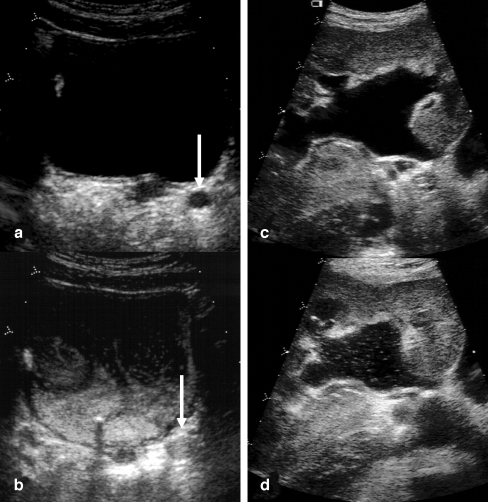
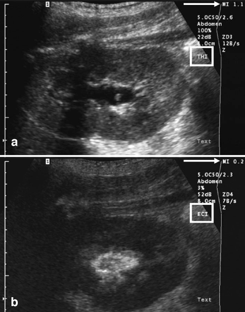
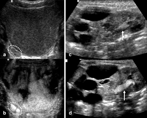
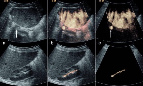
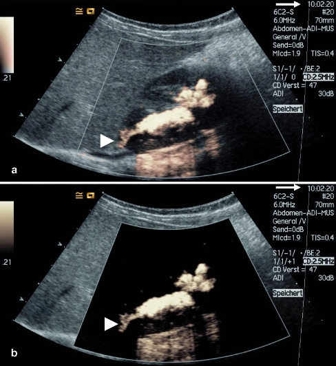
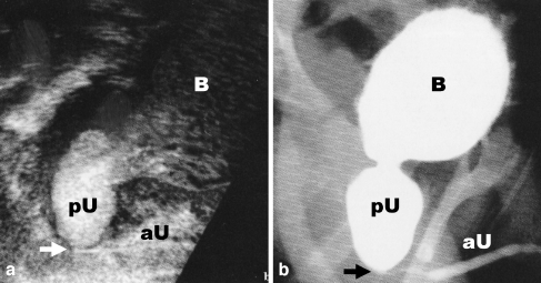
Voiding urosonography (VUS) encompasses examination of the urinary tract with intravesical administration of US contrast agent (UCA) for diagnosis of vesicoureteric reflux (VUR). The real breakthrough for US examination of VUR came with the availability of stabilized UCAs in the mid-1990s. This article presents a comprehensive review of various procedural aspects of VUS. Different US modalities are available for detecting the echogenic microbubbles: fundamental mode, colour Doppler US, harmonic imaging and dedicated contrast imaging with multiple display options. The reflux is graded (1 to 5) in a similar manner to the system used in voiding cystourethrography (VCUG). The most commonly used UCA for VUS, Levovist, is galactose-based and contains air-filled microbubbles. The recommended concentration is 300 mg/ml at a dose of 5-10%, or less than 5%, of the bladder filling volume when using fundamental or harmonic imaging modes, respectively. There are preliminary reports of VUS using a second-generation UCA, SonoVue. Here the UCA volume is less than 1% of the bladder filling volume. There is no specific contraindication to intravesical administration of UCA. The safety profile of intravesical Levovist is very high with no reports of side effects over a decade of use in VUS.
排尿期超声造影(VUS)包括经膀胱内注入超声造影剂(UCA)对尿路进行检查,以诊断膀胱输尿管反流(VUR)。20世纪90年代中期稳定型UCA的出现为超声检查VUR带来了真正的突破。本文对VUS的各个操作环节进行了全面综述。有多种超声模式可用于检测回声微泡:基波模式、彩色多普勒超声、谐波成像以及具有多种显示选项的专用造影成像。反流分级为(1至5级),与排尿性膀胱尿道造影(VCUG)所采用的系统分级方式类似。VUS最常用的UCA是声诺维,它是以半乳糖为基础的,含有充气微泡。当分别使用基波或谐波成像模式时,推荐浓度为300mg/ml,剂量分别为膀胱充盈量 的5 - 10%或小于5%。有关于使用第二代UCA声诺维进行VUS的初步报告。此时UCA的体积小于膀胱充盈量的1%。膀胱内注入UCA没有特定的禁忌证。膀胱内使用声诺维的安全性非常高,在VUS中使用十多年来没有副作用报告。