Gu Ping, Harwood Laura J, Zhang Xiaohong, Wylie Mildred, Curry W James, Cogliati Tiziana
Centre for Vision Sciences, Queens University Belfast, United Kingdom.
Mol Vis. 2007 Jun 29;13:1045-57.
Retinal progenitor cells (RPCs) and retinal stem cells (RSCs) from rodents and humans have been isolated and characterized in vitro. Transplantation experiments have confirmed their potential as tools for cell replacement in retinal degenerative diseases. The pig represents an ideal pre-clinical animal model to study the impact of transplantation because of the similarity of its eye to the human eye. However, little is known about porcine RPCs and RSCs. We aimed to identify and characterize in vitro RPCs and RSCs from porcine ocular tissues.
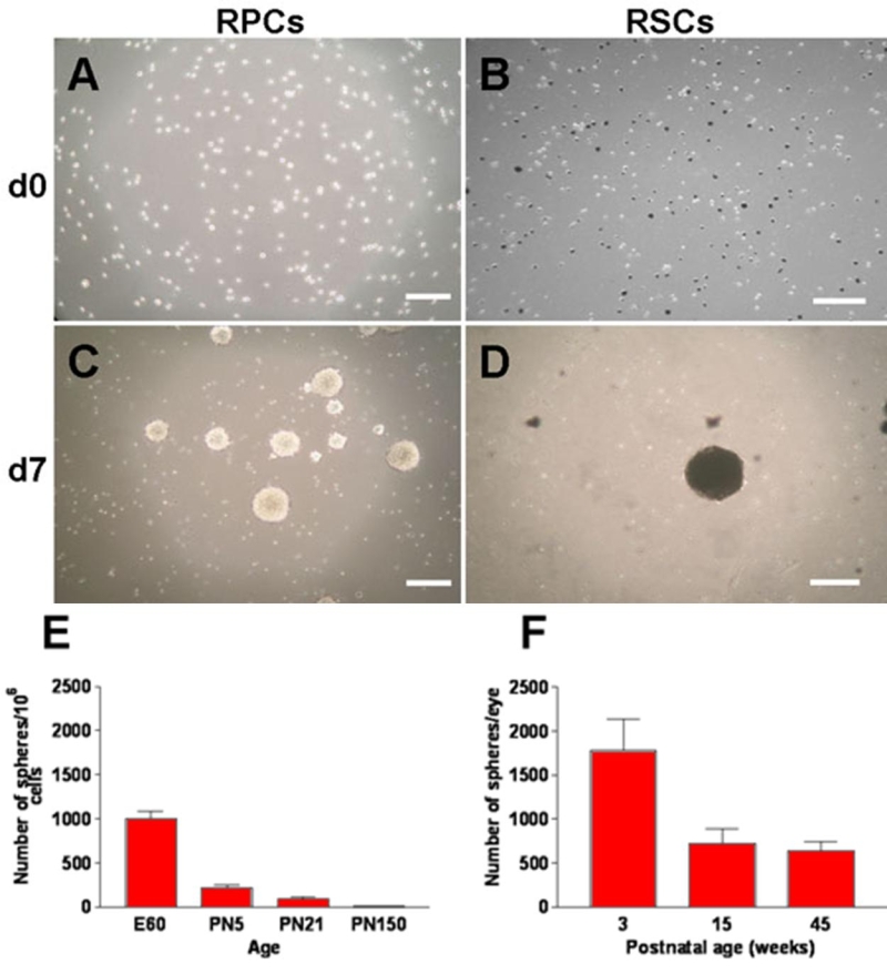
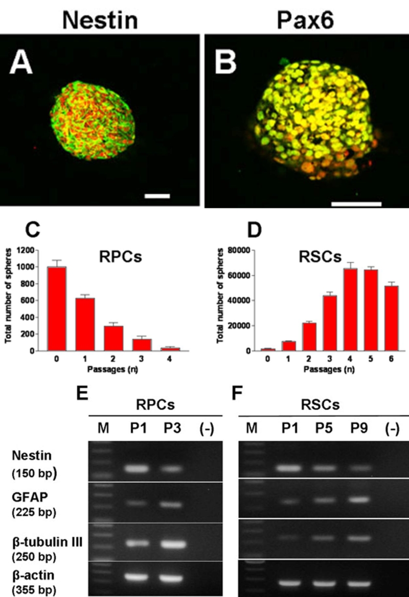
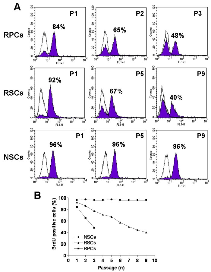
Cells from different subregions of embryonic, postnatal and adult porcine eyes were grown in suspension sphere culture in serum-free medium containing basic fibroblast growth factor (bFGF) and epidermal growth factor (EGF). Growth curves and BrdU incorporation assays were performed to establish the proliferative capacity of isolated porcine retina-derived RPCs and ciliary epithelium (CE)-derived RSCs. Self-renewal potential was investigated by subsphere formation assays. Changes in gene expression were assayed by reverse transcription polymerase chain reaction (RT-PCR) at different passages in culture. Finally, differentiation was induced by addition of serum to the cultures and expression of markers for retinal cell types was detected by immunohistochemical staining with specific antibodies.
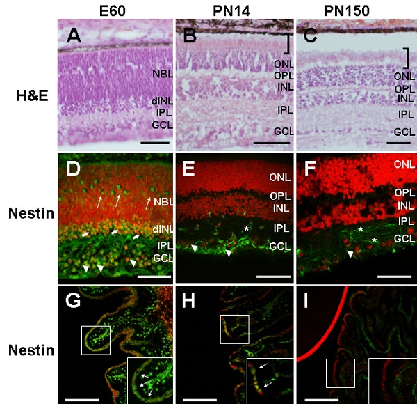
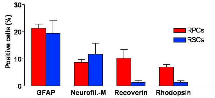
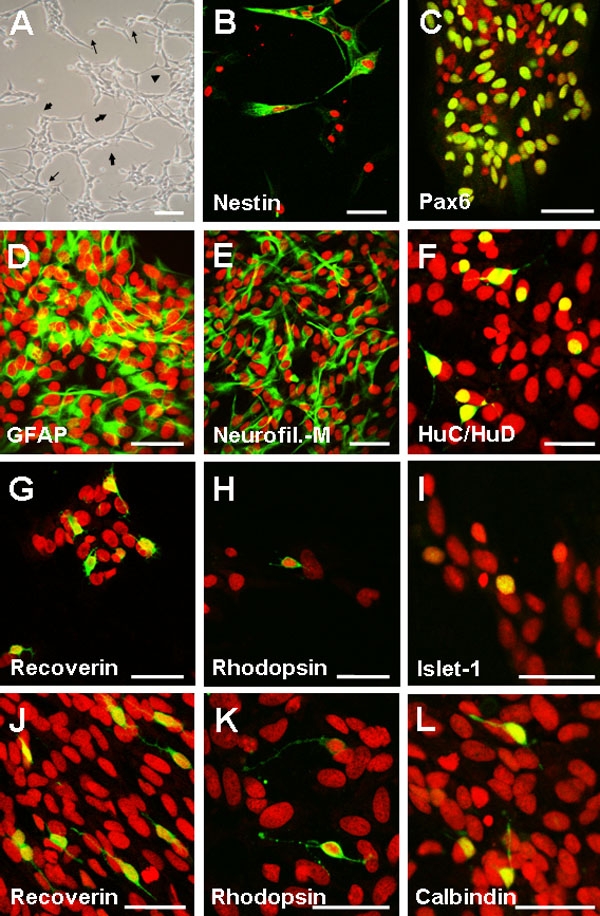
Dissociated cells from embryonic retina and CE at different postnatal ages generated primary nestin- and Pax6-immunoreactive neurosphere colonies in vitro in numbers that decreased with age. Embryonic and postnatal retina-derived RPCs and young CE-derived RSCs displayed self-renewal capacity, generating secondary neurosphere colonies. However, their self-renewal and proliferation capacity gradually decreased and they became more committed to differentiated states with subsequent passages. The expansion capacity of RPCs and RSCs was higher when they were maintained in monolayer culture. Porcine RPCs and RSCs could be induced to differentiate in vitro to express markers of retinal neurons and glia.
Porcine retina and CE contain RPCs and RSCs which are undifferentiated, self-renewing and multipotent and which show characteristics similar to their human counterparts. Therefore, the pig could be a useful source of cells to further investigate the cell biology of RPCs and RSCs and it could be used as a non-primate large animal model for pre-clinical studies on stem cell-based approaches to regenerative medicine in the retina.
已在体外分离并鉴定了来自啮齿动物和人类的视网膜祖细胞(RPCs)和视网膜干细胞(RSCs)。移植实验证实了它们作为视网膜退行性疾病细胞替代工具的潜力。由于猪眼与人眼相似,猪是研究移植影响的理想临床前动物模型。然而,关于猪RPCs和RSCs的了解甚少。我们旨在从猪眼组织中体外鉴定和表征RPCs和RSCs。
将来自胚胎、出生后和成年猪眼不同亚区域的细胞在含有碱性成纤维细胞生长因子(bFGF)和表皮生长因子(EGF)的无血清培养基中进行悬浮球培养。进行生长曲线和BrdU掺入试验以确定分离的猪视网膜来源的RPCs和睫状体上皮(CE)来源的RSCs的增殖能力。通过亚球形成试验研究自我更新潜力。在培养的不同传代时通过逆转录聚合酶链反应(RT-PCR)测定基因表达的变化。最后,通过向培养物中添加血清诱导分化,并用特异性抗体进行免疫组织化学染色检测视网膜细胞类型标志物的表达。
来自胚胎视网膜和不同出生后年龄的CE的解离细胞在体外产生了原代巢蛋白和Pax6免疫反应性神经球集落,其数量随年龄减少。胚胎和出生后视网膜来源的RPCs以及年轻CE来源的RSCs表现出自我更新能力,产生次级神经球集落。然而,它们的自我更新和增殖能力逐渐下降,并且随着后续传代它们更倾向于分化状态。当RPCs和RSCs维持在单层培养时,其扩增能力更高。猪RPCs和RSCs可在体外被诱导分化以表达视网膜神经元和神经胶质细胞的标志物。
猪视网膜和CE含有未分化、自我更新且具有多能性的RPCs和RSCs,其表现出与人类对应细胞相似的特征。因此,猪可能是进一步研究RPCs和RSCs细胞生物学的有用细胞来源,并且可作为非灵长类大型动物模型用于基于干细胞的视网膜再生医学临床前研究。