Scott Ingrid U, Edwards Allison R, Beck Roy W, Bressler Neil M, Chan Clement K, Elman Michael J, Friedman Scott M, Greven Craig Michael, Maturi Raj K, Pieramici Dante J, Shami Michel, Singerman Lawrence J, Stockdale Cynthia R
Jaeb Center for Health Research, 15310 Amberly Drive, Suite 350, Tampa, FL 33647, USA.
Ophthalmology. 2007 Oct;114(10):1860-7. doi: 10.1016/j.ophtha.2007.05.062. Epub 2007 Aug 15.
To provide data on the short-term effect of intravitreal bevacizumab for diabetic macular edema (DME).
Randomized phase II clinical trial.
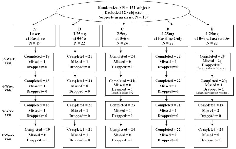
One hundred twenty-one eyes of 121 subjects (109 eligible for analysis) with DME and Snellen acuity equivalent ranging from 20/32 to 20/320.
Random assignment to 1 of 5 groups: (A) focal photocoagulation at baseline (n = 19), (B) intravitreal injection of 1.25 mg of bevacizumab at baseline and 6 weeks (n = 22), (C) intravitreal injection of 2.5 mg of bevacizumab at baseline and 6 weeks (n = 24), (D) intravitreal injection of 1.25 mg of bevacizumab at baseline and sham injection at 6 weeks (n = 22), or (E) intravitreal injection of 1.25 mg of bevacizumab at baseline and 6 weeks with photocoagulation at 3 weeks (n = 22).
Central subfield thickness (CST) on optical coherence tomography and best-corrected visual acuity (VA) were measured at baseline and after 3, 6, 9, 12, 18, and 24 weeks.
At baseline, median CST was 411 mum and median Snellen VA equivalent was 20/50. Compared with group A, groups B and C had a greater reduction in CST at 3 weeks and about 1 line better median VA over 12 weeks. There were no meaningful differences between groups B and C in CST reduction or VA improvement. A CST reduction > 11% (reliability limit) was present at 3 weeks in 36 of 84 (43%) bevacizumab-treated eyes and 5 of 18 (28%) eyes treated with laser alone, and at 6 weeks in 31 of 84 (37%) and 9 of 18 (50%) eyes, respectively. Combining focal photocoagulation with bevacizumab resulted in no apparent short-term benefit or adverse outcomes. Endophthalmitis developed in 1 eye. The following events occurred during the first 24 weeks in subjects treated with bevacizumab without attributing cause to the drug: myocardial infarction (n = 2), congestive heart failure (n = 1), elevated blood pressure (n = 3), and worsened renal function (n = 3).
These results demonstrate that intravitreal bevacizumab can reduce DME in some eyes, but the study was not designed to determine whether treatment is beneficial. A phase III trial would be needed for that purpose.
提供玻璃体内注射贝伐单抗治疗糖尿病性黄斑水肿(DME)的短期疗效数据。
随机II期临床试验。
121例受试者的121只眼(109例符合分析条件)患有DME,Snellen视力相当于20/32至20/320。
随机分为5组中的1组:(A)基线时进行局部光凝(n = 19),(B)基线和6周时玻璃体内注射1.25 mg贝伐单抗(n = 22),(C)基线和6周时玻璃体内注射2.5 mg贝伐单抗(n = 24),(D)基线时玻璃体内注射1.25 mg贝伐单抗,6周时进行假注射(n = 22),或(E)基线和6周时玻璃体内注射1.25 mg贝伐单抗,3周时进行光凝(n = 22)。
在基线以及3、6、9、12、18和24周后,通过光学相干断层扫描测量中心子野厚度(CST),并测量最佳矫正视力(VA)。
基线时,CST中位数为411μm,Snellen视力相当于20/50。与A组相比,B组和C组在3周时CST降低幅度更大,在12周内中位数VA提高约1行。B组和C组在CST降低或VA改善方面无显著差异。在3周时,84只接受贝伐单抗治疗的眼中有36只(43%)和仅接受激光治疗的18只眼中有5只(28%)CST降低>11%(可靠限度);在6周时,分别为84只眼中的31只(37%)和18只眼中的9只(50%)。将局部光凝与贝伐单抗联合使用未产生明显的短期益处或不良后果。1只眼发生了眼内炎。在接受贝伐单抗治疗的受试者的前24周内发生了以下事件,但未将原因归因于药物:心肌梗死(n = 2)、充血性心力衰竭(n = 1)、血压升高(n = 3)和肾功能恶化(n = 3)。
这些结果表明玻璃体内注射贝伐单抗可使部分眼的DME减轻,但该研究并非旨在确定治疗是否有益。为此需要进行III期试验。