Li Zongjin, Wu Jenny C, Sheikh Ahmad Y, Kraft Daniel, Cao Feng, Xie Xiaoyan, Patel Manishkumar, Gambhir Sanjiv S, Robbins Robert C, Cooke John P, Wu Joseph C
Department of Radiology, Stanford University School of Medicine, Stanford, CA 94305, USA.
Circulation. 2007 Sep 11;116(11 Suppl):I46-54. doi: 10.1161/CIRCULATIONAHA.106.680561.
Embryonic stem (ES) cells are distinguished by their capacity for self-renewal and pluripotency. Here we characterize the differentiation of ES cell-derived endothelial cells (ESC-ECs), use molecular imaging techniques to examine their survival in vivo, and determine the therapeutic efficacy of ESC-ECs for restoration of cardiac function after ischemic injury.
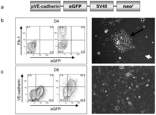
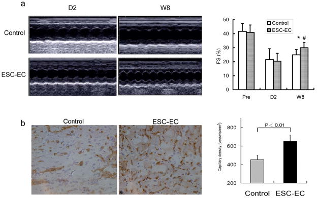
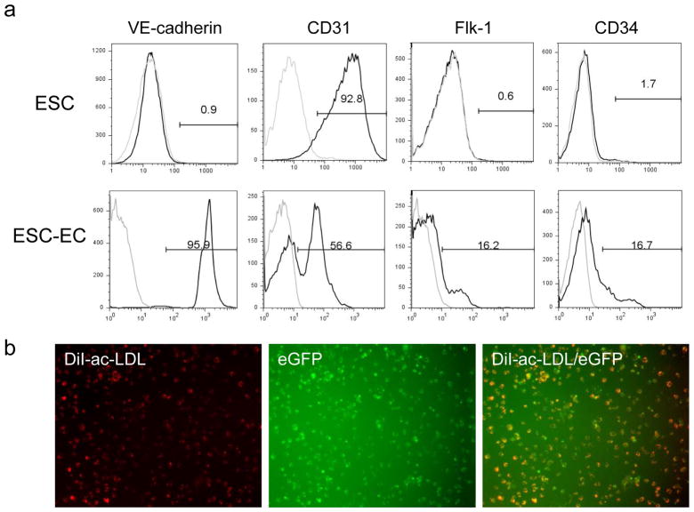
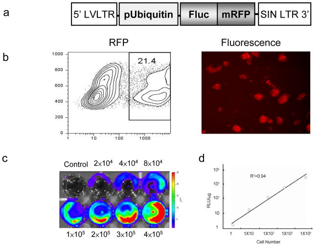
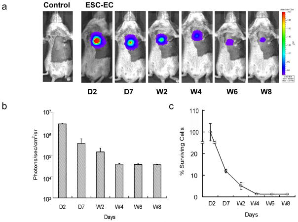
Murine ES cells were transfected with a construct composed of a vascular endothelial cadherin promoter driving enhanced green fluorescence protein (pVE-cadherin-eGFP). Differentiation of ES cells to ECs was detected by FACS analysis using Flk-1 (early EC marker at day 4) and VE-cadherin (late EC marker at day 8). After isolation, these ESC-ECs express endothelial cell markers similar to adult mouse lung endothelial cells, form vascular-like channels, and incorporate DiI-labeled acetylated low-density lipoprotein (DiI-Ac-LDL). For in vivo imaging, ES cells were transduced with an ubiquitin promoter driving firefly luciferase and monomeric red fluorescence protein (pUb-Fluc-mRFP). A robust correlation exists between Fluc signals and cell numbers by ex vivo imaging analysis (R2=0.98) and by in vitro enzyme assay (R2=0.94). Afterward, 5x10(5) ESC-ECs or PBS (as control) was injected into the hearts of mice undergoing LAD ligation (n=15 per group). Bioluminescence imaging showed longitudinal survival of transplanted ESC-ECs for approximately 8 weeks. Echocardiogram demonstrated significant functional improvement in the ESC-EC group compared with control (P=0.04). Finally, postmortem analysis confirmed increased presence of small capillaries and venules in the infarcted zones by CD31 staining.
This is the first study to track the fate and function of transplanted ESC-ECs in the heart. With further validation, these ESC-ECs could become a valuable source of cell therapy for induction of angiogenesis in the treatment of myocardial ischemia.
胚胎干细胞(ES细胞)以其自我更新能力和多能性而著称。在此,我们对ES细胞来源的内皮细胞(ESC-ECs)的分化进行了表征,运用分子成像技术检测它们在体内的存活情况,并确定ESC-ECs对缺血性损伤后心脏功能恢复的治疗效果。
用一个由血管内皮钙黏蛋白启动子驱动增强型绿色荧光蛋白的构建体(pVE-钙黏蛋白-eGFP)转染小鼠ES细胞。使用Flk-1(第4天的早期内皮细胞标志物)和VE-钙黏蛋白(第8天的晚期内皮细胞标志物)通过流式细胞术分析检测ES细胞向内皮细胞的分化。分离后,这些ESC-ECs表达与成年小鼠肺内皮细胞相似的内皮细胞标志物,形成血管样通道,并摄取DiI标记的乙酰化低密度脂蛋白(DiI-Ac-LDL)。为了进行体内成像,用一个驱动萤火虫荧光素酶和单体红色荧光蛋白的泛素启动子(pUb-Fluc-mRFP)转导ES细胞。通过离体成像分析(R2 = 0.98)和体外酶测定(R2 = 0.94),Fluc信号与细胞数量之间存在很强的相关性。之后,将5×10(5)个ESC-ECs或PBS(作为对照)注射到接受左前降支结扎的小鼠心脏中(每组n = 15)。生物发光成像显示移植的ESC-ECs纵向存活约8周。超声心动图显示ESC-EC组与对照组相比有显著的功能改善(P = 0.04)。最后,尸检分析通过CD31染色证实梗死区域中小毛细血管和小静脉的存在增加。
这是第一项追踪移植到心脏中的ESC-ECs的命运和功能的研究。经过进一步验证,这些ESC-ECs可能成为细胞治疗中诱导血管生成以治疗心肌缺血的宝贵细胞来源。