Division of Cardiovascular Medicine, Stanford University School of Medicine, Stanford, CA, USA.
Arterioscler Thromb Vasc Biol. 2011 Nov;31(11):e72-9. doi: 10.1161/ATVBAHA.111.230938.
Stem cell therapy for angiogenesis and vascular regeneration has been investigated using adult or embryonic stem cells. In the present study, we investigated the potential of endothelial cells (ECs) derived from human induced pluripotent stem cells (hiPSCs) to promote the perfusion of ischemic tissue in a murine model of peripheral arterial disease.
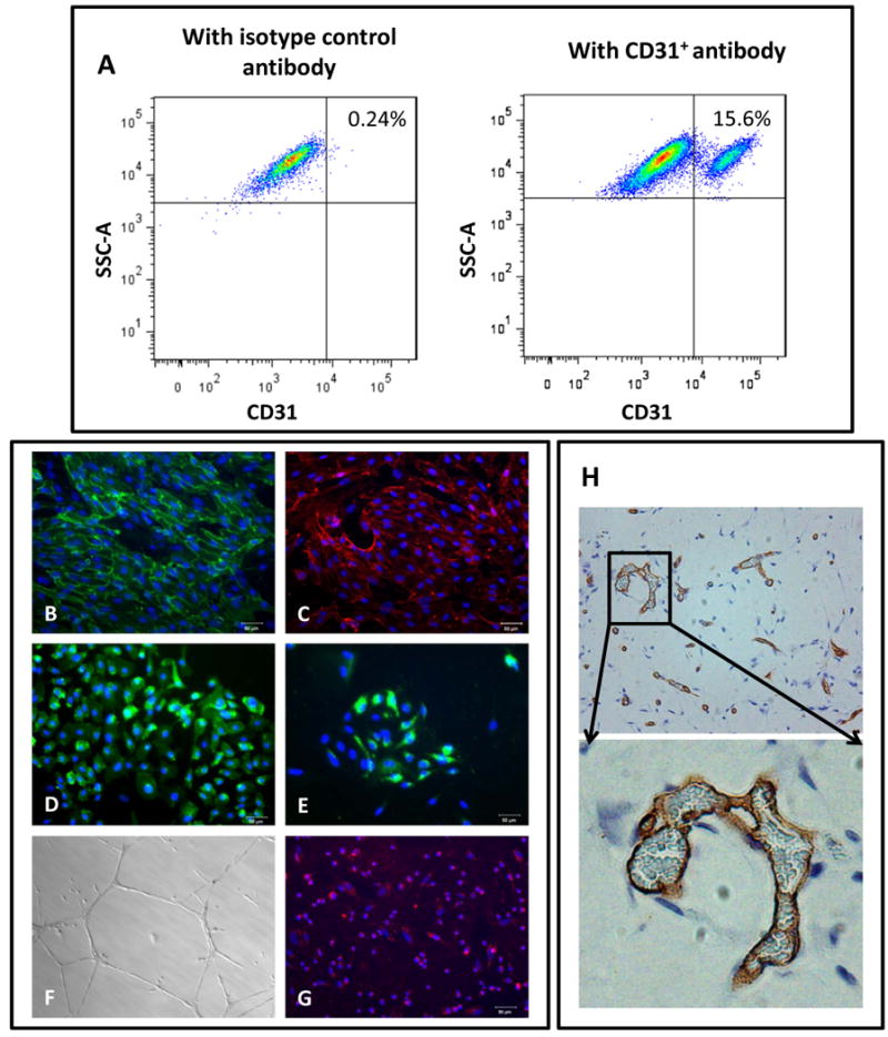
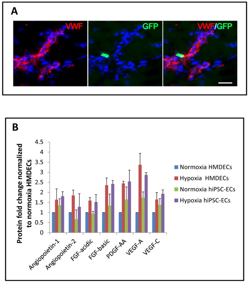
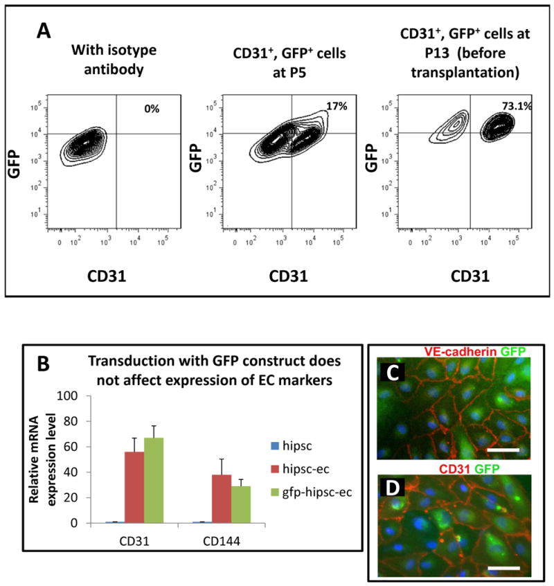
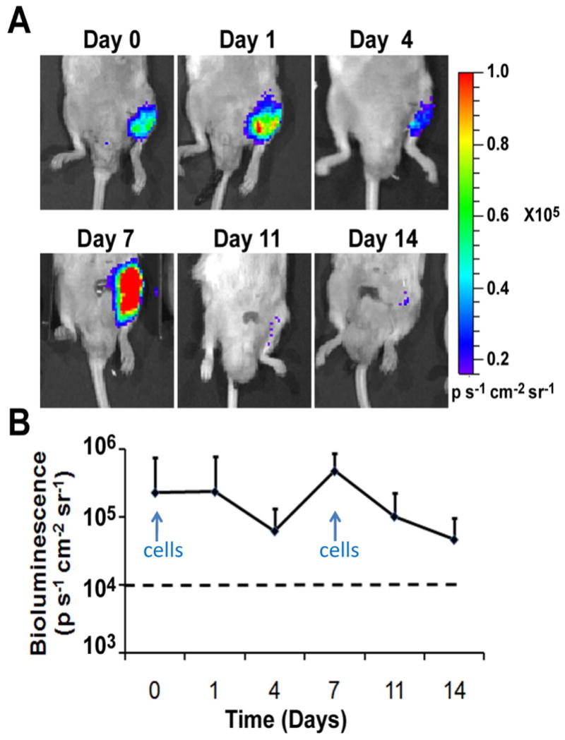
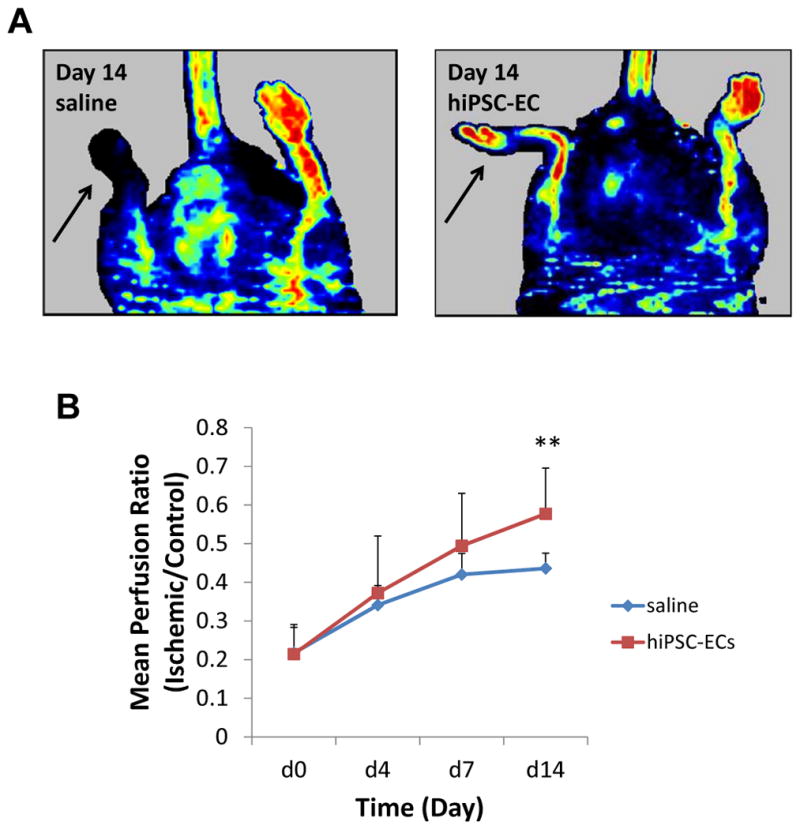
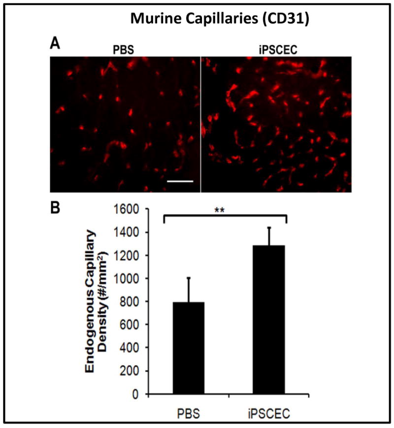
Endothelial differentiation was initiated by culturing hiPSCs for 14 days in differentiation media supplemented with BMP-4 and vascular endothelial growth factor. The hiPSC-ECs exhibited endothelial characteristics by forming capillary-like structures in matrigel and incorporating acetylated-LDL. They stained positively for EC markers such as KDR, CD31, CD144, and eNOS. In vitro exposure of hiPSC-ECs to hypoxia resulted in increased expression of various angiogenic related cytokines and growth factors. hiPSC-ECs were stably transduced with a double fusion construct encoded by the ubiquitin promoter, firefly luciferase for bioluminescence imaging and green fluorescence protein for fluorescent detection. The hiPSC-ECs (5×10(5)) were delivered by intramuscular injection into the ischemic hindlimb of SCID mice at day 0 and again on day 7 after femoral artery ligation (n=8). Bioluminescence imaging showed that hiPSC-ECs survived in the ischemic limb for at least 2 weeks. In addition, laser Doppler imaging showed that the ratio of blood perfusion was increased by hiPSC-EC treatment by comparison to the saline-treated group (0.58±0.12 versus 0.44±0.04; P=0.005). The total number of capillaries in the ischemic limb of mice receiving hiPSC-EC injections was greater than those in the saline-treated group (1284±155 versus 797±206 capillaries/mm(2)) (P<0.002).
This study is a first step toward development of a regenerative strategy for peripheral arterial disease based on the use of ECs derived from hiPSCs.
使用成体或胚胎干细胞对血管生成和血管再生的干细胞治疗进行了研究。在本研究中,我们研究了源自人诱导多能干细胞(hiPSC)的内皮细胞(EC)在小鼠外周动脉疾病模型中促进缺血组织灌注的潜力。
通过在补充有 BMP-4 和血管内皮生长因子的分化培养基中培养 hiPSC 14 天来启动内皮分化。hiPSC-EC 在 Matrigel 中形成类似毛细血管的结构并摄取乙酰化 LDL,从而表现出内皮细胞特征。它们对 EC 标志物如 KDR、CD31、CD144 和 eNOS 呈阳性染色。hiPSC-EC 在体外暴露于低氧条件下导致各种血管生成相关细胞因子和生长因子的表达增加。用由泛素启动子、萤火虫荧光素酶编码的双融合构建体稳定转导 hiPSC-EC 用于生物发光成像和绿色荧光蛋白用于荧光检测。在股动脉结扎后第 0 天和第 7 天(n=8)将 hiPSC-EC(5×10(5))通过肌肉内注射递送至缺血后肢。生物发光成像显示 hiPSC-EC 在缺血肢体中至少存活 2 周。此外,激光多普勒成像显示 hiPSC-EC 治疗使与盐水处理组相比,血液灌注比增加(0.58±0.12 对 0.44±0.04;P=0.005)。接受 hiPSC-EC 注射的小鼠缺血肢体中的毛细血管总数大于盐水处理组(1284±155 对 797±206 毛细血管/mm(2))(P<0.002)。
这项研究是基于使用 hiPSC 衍生的 EC 开发外周动脉疾病再生策略的第一步。