• 文献检索
  • 文档翻译
  • 深度研究
  • 学术资讯
  • Suppr Zotero 插件Zotero 插件
  • 邀请有礼
  • 套餐&价格
  • 历史记录
应用&插件
Suppr Zotero 插件Zotero 插件浏览器插件Mac 客户端Windows 客户端微信小程序
定价
高级版会员购买积分包购买API积分包
服务
文献检索文档翻译深度研究API 文档MCP 服务
关于我们
关于 Suppr公司介绍联系我们用户协议隐私条款
关注我们

Suppr 超能文献

核心技术专利:CN118964589B侵权必究
粤ICP备2023148730 号-1Suppr @ 2026

文献检索

告别复杂PubMed语法,用中文像聊天一样搜索,搜遍4000万医学文献。AI智能推荐,让科研检索更轻松。

立即免费搜索

文件翻译

保留排版,准确专业,支持PDF/Word/PPT等文件格式,支持 12+语言互译。

免费翻译文档

深度研究

AI帮你快速写综述,25分钟生成高质量综述,智能提取关键信息,辅助科研写作。

立即免费体验

创建大脑和颅骨的物理3D立体光刻模型。

Creating physical 3D stereolithograph models of brain and skull.

作者信息

Kelley Daniel J, Farhoud Mohammed, Meyerand M Elizabeth, Nelson David L, Ramirez Lincoln F, Dempsey Robert J, Wolf Alan J, Alexander Andrew L, Davidson Richard J

机构信息

Waisman Laboratory for Brain Imaging and Behavior, Waisman Center, Madison, Wisconsin, United States of America.

出版信息

PLoS One. 2007 Oct 31;2(10):e1119. doi: 10.1371/journal.pone.0001119.

DOI:10.1371/journal.pone.0001119
PMID:17971879
原文链接:https://pmc.ncbi.nlm.nih.gov/articles/PMC2040197/
Abstract

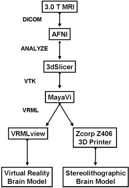
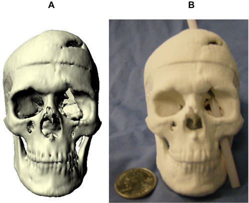
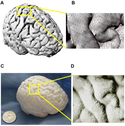
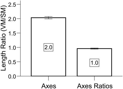
The human brain and skull are three dimensional (3D) anatomical structures with complex surfaces. However, medical images are often two dimensional (2D) and provide incomplete visualization of structural morphology. To overcome this loss in dimension, we developed and validated a freely available, semi-automated pathway to build 3D virtual reality (VR) and hand-held, stereolithograph models. To evaluate whether surface visualization in 3D was more informative than in 2D, undergraduate students (n = 50) used the Gillespie scale to rate 3D VR and physical models of both a living patient-volunteer's brain and the skull of Phineas Gage, a historically famous railroad worker whose misfortune with a projectile tamping iron provided the first evidence of a structure-function relationship in brain. Using our processing pathway, we successfully fabricated human brain and skull replicas and validated that the stereolithograph model preserved the scale of the VR model. Based on the Gillespie ratings, students indicated that the biological utility and quality of visual information at the surface of VR and stereolithograph models were greater than the 2D images from which they were derived. The method we developed is useful to create VR and stereolithograph 3D models from medical images and can be used to model hard or soft tissue in living or preserved specimens. Compared to 2D images, VR and stereolithograph models provide an extra dimension that enhances both the quality of visual information and utility of surface visualization in neuroscience and medicine.

摘要

人类大脑和颅骨是具有复杂表面的三维(3D)解剖结构。然而,医学图像通常是二维(2D)的,无法完整呈现结构形态。为了克服这种维度上的缺失,我们开发并验证了一种免费的半自动方法,用于构建3D虚拟现实(VR)模型和手持式立体光刻模型。为了评估3D表面可视化是否比2D更具信息性,我们让50名本科生使用吉莱斯皮量表,对一名活体患者志愿者的大脑以及历史上著名的铁路工人菲尼亚斯·盖奇的颅骨的3D VR模型和实体模型进行评分。盖奇曾不幸被射弹夯铁击中,这一事件首次证明了大脑中结构与功能的关系。通过我们的处理方法,我们成功制作了人类大脑和颅骨复制品,并验证了立体光刻模型保留了VR模型的比例。根据吉莱斯皮评分,学生们表示,VR模型和立体光刻模型表面视觉信息的生物学效用和质量高于其所源自的2D图像。我们开发的方法有助于从医学图像创建VR和立体光刻3D模型,可用于对活体或保存标本中的硬组织或软组织进行建模。与2D图像相比,VR和立体光刻模型提供了一个额外的维度,提高了神经科学和医学中视觉信息的质量以及表面可视化的效用。

https://cdn.ncbi.nlm.nih.gov/pmc/blobs/8382/2040197/b9c349213460/pone.0001119.g004.jpg
https://cdn.ncbi.nlm.nih.gov/pmc/blobs/8382/2040197/eb767be98a95/pone.0001119.g001.jpg
https://cdn.ncbi.nlm.nih.gov/pmc/blobs/8382/2040197/e4adb19f6d13/pone.0001119.g002.jpg
https://cdn.ncbi.nlm.nih.gov/pmc/blobs/8382/2040197/f84b76a0c788/pone.0001119.g003.jpg
https://cdn.ncbi.nlm.nih.gov/pmc/blobs/8382/2040197/b9c349213460/pone.0001119.g004.jpg

相似文献

1
Creating physical 3D stereolithograph models of brain and skull.创建大脑和颅骨的物理3D立体光刻模型。
PLoS One. 2007 Oct 31;2(10):e1119. doi: 10.1371/journal.pone.0001119.
2
Creating 3D models from Radiologic Images for Virtual Reality Medical Education Modules.从放射影像创建 3D 模型,用于虚拟现实医学教育模块。
J Med Syst. 2019 May 3;43(6):166. doi: 10.1007/s10916-019-1308-3.
3
Experimental evidence for improved neuroimaging interpretation using three-dimensional graphic models.使用三维图形模型提高神经影像学解读的实验证据。
Anat Sci Educ. 2012 May-Jun;5(3):132-7. doi: 10.1002/ase.1275. Epub 2012 Mar 20.
4
Skull stripping based on region growing for magnetic resonance brain images.基于区域生长的磁共振脑图像颅骨剥离
Neuroimage. 2009 Oct 1;47(4):1394-407. doi: 10.1016/j.neuroimage.2009.04.047. Epub 2009 Apr 21.
5
JAtlasView: a Java atlas-viewer for browsing biomedical 3D images and atlases.JAtlasView:一款用于浏览生物医学3D图像和图谱的Java图谱查看器。
BMC Bioinformatics. 2005 Mar 9;6:47. doi: 10.1186/1471-2105-6-47.
6
Surface modeling--uncertainty estimation and visualization.表面建模——不确定性估计与可视化
Comput Med Imaging Graph. 2008 Mar;32(2):134-9. doi: 10.1016/j.compmedimag.2007.10.006. Epub 2007 Dec 21.
7
Can virtual reality improve traditional anatomy education programmes? A mixed-methods study on the use of a 3D skull model.虚拟现实能否改善传统解剖学教育课程?一项关于使用3D颅骨模型的混合方法研究。
BMC Med Educ. 2020 Oct 31;20(1):395. doi: 10.1186/s12909-020-02255-6.
8
Three-Dimensional Modeling and Extended Reality Simulations of the Cross-Sectional Anatomy of the Cerebrum, Cerebellum, and Brainstem.大脑、小脑和脑干的横断解剖的三维建模和扩展现实模拟。
Oper Neurosurg (Hagerstown). 2023 Jul 1;25(1):3-10. doi: 10.1227/ons.0000000000000703. Epub 2023 Apr 21.
9
Display of fused images: methods, interpretation, and diagnostic improvements.融合图像的展示:方法、解读及诊断改进。
Semin Nucl Med. 2003 Jul;33(3):219-27. doi: 10.1053/snuc.2003.127311.
10
Skull-stripping with machine learning deformable organisms.使用机器学习可变形模型进行颅骨剥离
J Neurosci Methods. 2014 Oct 30;236:114-24. doi: 10.1016/j.jneumeth.2014.07.023. Epub 2014 Aug 12.

引用本文的文献

1
Neuroscience Education Begins With Good Science: Communication About Phineas Gage (1823-1860), One of Neurology's Most-Famous Patients, in Scientific Articles.神经科学教育始于严谨的科学:关于菲尼亚斯·盖奇(1823 - 1860)的科学文章中的交流,他是神经学领域最著名的患者之一。
Front Hum Neurosci. 2022 Apr 28;16:734174. doi: 10.3389/fnhum.2022.734174. eCollection 2022.
2
Three-Dimensional Printing: Custom-Made Implants for Craniomaxillofacial Reconstructive Surgery.三维打印:用于颅颌面重建手术的定制植入物
Craniomaxillofac Trauma Reconstr. 2017 Jun;10(2):89-98. doi: 10.1055/s-0036-1594277. Epub 2017 Jan 3.
3
Weight lifting can facilitate appreciative comprehension for museum exhibits.

本文引用的文献

1
Play dough as an educational tool for visualization of complicated cerebral aneurysm anatomy.橡皮泥作为一种用于可视化复杂脑动脉瘤解剖结构的教育工具。
BMC Med Educ. 2005 May 10;5(1):15. doi: 10.1186/1472-6920-5-15.
2
BrainTrain: brain simulator for medical VR application.BrainTrain:用于医学虚拟现实应用的大脑模拟器。
Stud Health Technol Inform. 2005;111:378-84.
3
Prospective study on cranioplasty with individual carbon fiber reinforced polymer (CFRP) implants produced by means of stereolithography.关于采用立体光刻技术制作的个体化碳纤维增强聚合物(CFRP)植入物进行颅骨修补术的前瞻性研究。
举重可以促进对博物馆展品的欣赏理解。
Front Psychol. 2014 Apr 14;5:307. doi: 10.3389/fpsyg.2014.00307. eCollection 2014.
4
A deformable generic 3D model of haptoral anchor of Monogenean.一种变形通用 3D 模型的 Monogenean 的固着器。
PLoS One. 2013 Oct 28;8(10):e77650. doi: 10.1371/journal.pone.0077650. eCollection 2013.
5
An imaging roadmap for biology education: from nanoparticles to whole organisms.生物学教育的成像路线图:从纳米颗粒到整个生物体。
CBE Life Sci Educ. 2008 Summer;7(2):202-9. doi: 10.1187/cbe.07-10-0094.
Surg Neurol. 2004 Dec;62(6):510-21. doi: 10.1016/j.surneu.2004.01.025.
4
Images in clinical medicine. The tale of Phineas Gage, digitally remastered.临床医学影像。菲尼亚斯·盖奇的故事,数字修复版。
N Engl J Med. 2004 Dec 2;351(23):e21. doi: 10.1056/NEJMicm031024.
5
Short communication: fused deposition models from CT scans.
Br J Radiol. 2004 Jun;77(918):504-7. doi: 10.1259/bjr/50012454.
6
Cerebrovascular stereolithographic biomodeling for aneurysm surgery. Technical note.用于动脉瘤手术的脑血管立体光刻生物建模。技术说明。
J Neurosurg. 2004 Jan;100(1):139-45. doi: 10.3171/jns.2004.100.1.0139.
7
Virtual dissection: a lesson from the 18th century.虚拟解剖:来自18世纪的一课。
Lancet. 2003 Dec 20;362(9401):2110-3. doi: 10.1016/S0140-6736(03)15114-8.
8
The application of rapid prototyping techniques in cranial reconstruction and preoperative planning in neurosurgery.快速成型技术在神经外科颅骨重建及术前规划中的应用。
J Craniofac Surg. 2003 Nov;14(6):899-914. doi: 10.1097/00001665-200311000-00014.
9
Leonardo da Vinci's contributions to neuroscience.列奥纳多·达·芬奇对神经科学的贡献。
Trends Neurosci. 2002 Apr;25(4):217-20. doi: 10.1016/s0166-2236(00)02121-4.
10
An integrated visualization system for surgical planning and guidance using image fusion and an open MR.一种使用图像融合和开放式磁共振成像进行手术规划与引导的集成可视化系统。
J Magn Reson Imaging. 2001 Jun;13(6):967-75. doi: 10.1002/jmri.1139.