Chu Winnie Cw, Ng Bobby Kw, Li Albert M, Lam Tsz-Ping, Lam Wynnie Wm, Cheng Jack Cy
Departments of Diagnostic Radiology and Organ Imaging, The Chinese University of Hong Kong, Prince of Wales Hospital, Shatin, Hong Kong, China.
J Orthop Surg Res. 2007 Nov 19;2:20. doi: 10.1186/1749-799X-2-20.
Restrictive impairment is the commonest reported pulmonary deficit in AIS, which improves following surgical operation. However, exact mechanism of how improvement is brought about is unknown. Dynamic fast breath-hold (BH)-MR imaging is a recent advance which provides direct quantitative visual assessment of pulmonary function. By using above technique, change in lung volume, chest wall and diaphragmatic motion in AIS patients before and six months after posterior spinal fusion surgery were measured.
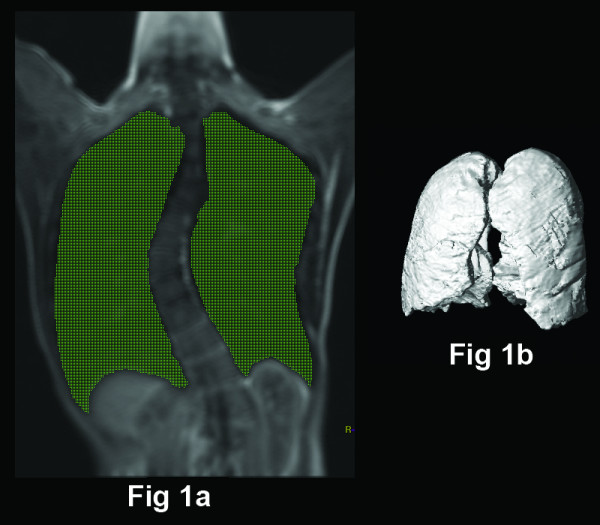
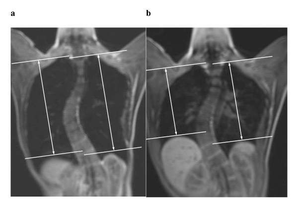
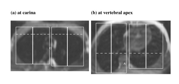
16 patients with severe right-sided predominant thoracic scoliosis (standing Cobb's angle 50 degrees -82 degrees , mean 60 degrees ) received posterior spinal fusion without thoracoplasty were recruited into this study. BH-MR sequences were used to obtain coronal images of the whole chest during full inspiration and expiration. The following measurements were assessed: (1) inspiratory, expiratory and change in lung volume; (2) change in anteroposterior (AP) and transverse (TS) diameter of the chest wall at two levels: carina and apex (3) change in diaphragmatic heights. The changes in parameters before and after operation were compared using Wilcoxon signed ranks test. Patients were also asked to score their breathing effort before and after operation using a scale of 1-9 with ascending order of effort. The degree of spinal surgical correction at three planes was also assessed by reformatted MR images and correction rate of Cobb's angle was calculated.
The individual or total inspiratory and expiratory volume showed slight but insignificant increase after operation. There was significantly increase in bilateral TS chest wall movement at carina level and increase in bilateral diaphragmatic movements between inspiration and expiration. The AP chest wall movements, however, did not significantly change.The median breathing effort after operation was lower than that before operation (p < 0.05).There was significant reduction in coronal Cobb's angle after operation but the change in sagittal and axial angle at scoliosis apex was not significant.
There is improvement of lateral chest wall and diaphragmatic motions in AIS patients six months after posterior spinal fusion, associated with subjective symptomatic improvement. Lung volumes however, do not significantly change after operation. BH-MR is novel non-invasive method for long term post operative assessment of pulmonary function in AIS patients.
限制性肺功能障碍是报道中青少年特发性脊柱侧弯(AIS)最常见的肺部缺陷,手术治疗后可改善。然而,改善是如何实现的确切机制尚不清楚。动态快速屏气(BH)-磁共振成像(MR)是一项最新进展,可对肺功能进行直接定量可视化评估。利用上述技术,测量了AIS患者后路脊柱融合手术前后及术后六个月肺容积、胸壁和膈肌运动的变化。
16例严重右侧为主的胸段脊柱侧弯患者(站立位Cobb角50度-82度,平均60度)接受了不进行胸廓成形术的后路脊柱融合术,并纳入本研究。BH-MR序列用于在全吸气和呼气时获取全胸的冠状位图像。评估以下测量指标:(1)吸气、呼气和肺容积变化;(2)在两个层面(隆突和顶点)胸壁前后径(AP)和横径(TS)的变化;(3)膈肌高度变化。采用Wilcoxon符号秩检验比较手术前后参数的变化。还要求患者使用1-9级评分量表(努力程度递增)对手术前后的呼吸努力程度进行评分。通过重新格式化的MR图像评估脊柱手术在三个平面上的矫正程度,并计算Cobb角的矫正率。
术后个体或总的吸气和呼气容积略有增加,但无统计学意义。隆突水平双侧胸壁TS运动显著增加,吸气和呼气之间双侧膈肌运动增加。然而,胸壁AP运动无显著变化。术后呼吸努力程度中位数低于术前(p<0.05)。术后冠状位Cobb角显著减小,但脊柱侧弯顶点矢状位和轴位角度变化不显著。
AIS患者后路脊柱融合术后六个月,胸壁外侧和膈肌运动得到改善,伴有主观症状改善。然而,术后肺容积无显著变化。BH-MR是一种用于AIS患者术后长期肺功能评估的新型无创方法。