• 文献检索
  • 文档翻译
  • 深度研究
  • 学术资讯
  • Suppr Zotero 插件Zotero 插件
  • 邀请有礼
  • 套餐&价格
  • 历史记录
应用&插件
Suppr Zotero 插件Zotero 插件浏览器插件Mac 客户端Windows 客户端微信小程序
定价
高级版会员购买积分包购买API积分包
服务
文献检索文档翻译深度研究API 文档MCP 服务
关于我们
关于 Suppr公司介绍联系我们用户协议隐私条款
关注我们

Suppr 超能文献

核心技术专利:CN118964589B侵权必究
粤ICP备2023148730 号-1Suppr @ 2026

文献检索

告别复杂PubMed语法,用中文像聊天一样搜索,搜遍4000万医学文献。AI智能推荐,让科研检索更轻松。

立即免费搜索

文件翻译

保留排版,准确专业,支持PDF/Word/PPT等文件格式,支持 12+语言互译。

免费翻译文档

深度研究

AI帮你快速写综述,25分钟生成高质量综述,智能提取关键信息,辅助科研写作。

立即免费体验

大鼠下颌第一磨牙牙板的发育与命运

The development and fate of the dental lamina of the mandibular first molar tooth in the rat.

作者信息

Khaejornbut J, Wilson D J, Owens P D

机构信息

Department of Oral Biology, Prince of Songkla University, Haadyai, Thailand.

出版信息

J Anat. 1991 Dec;179:85-96.

PMID:1817145
原文链接:https://pmc.ncbi.nlm.nih.gov/articles/PMC1260578/
Abstract

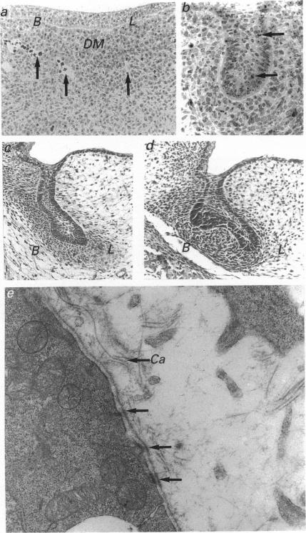
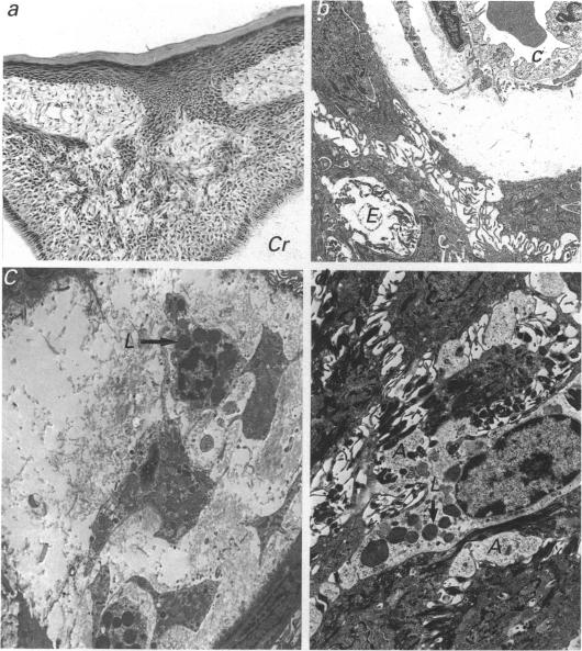
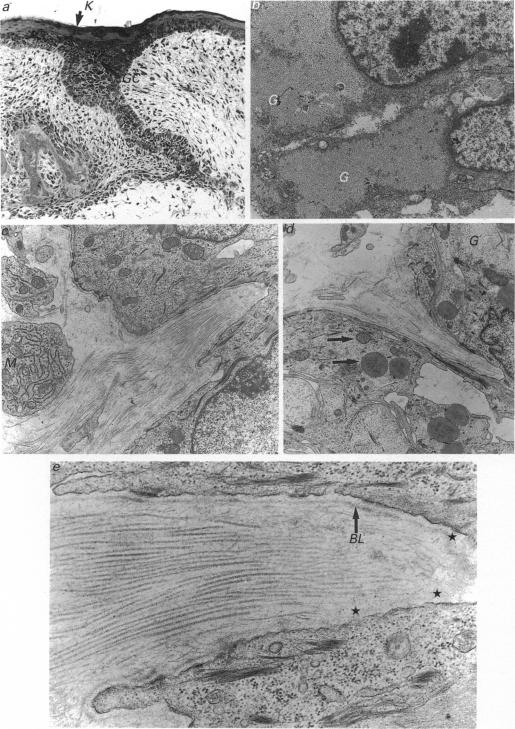
The lamina of the first mandibular molar teeth of rats, age range 13 d intrauterine (i.u.) to 16 d postnatal (p.n.), was examined by light and transmission electron microscopy to establish histological baselines of its development and fate. All material was obtained from animals anaesthetised with ether, killed by cervical dislocation and prepared by routine methods for both types of examination. Contrary to earlier reports that the lamina remains intact throughout development, mesenchymal elements disrupt the lamina. These were seen first at 19 d i.u., as collagen-filled bays in the basal epithelial layers, associated with partial loss of related basal lamina. In the early stages, collagen deposition was limited and it was not obviously preceded by epithelial cell death or transformation, even though many bay-related cells showed lipid and glycogen accumulations. Later disruption of the lamina showed more mesenchymal cells as well as collagen in deeper spaces. After the onset of tooth eruption, mesenchymal cells external to and within the lamina contained lysosomal bodies and these plus evidence of related epithelial cell death and capillaries in the laminar spaces became more and more apparent. Similar collagen deposits were observed in a successional tooth primordium, which appeared at term but eventually aborted between days 5 and 10 p.n. Thus disruption of the lamina by connective tissue began earlier than has been reported previously and progressed as the tooth erupted towards the oral cavity. The evidence suggests that this disruption is initiated and sustained by mesenchymal cell activity rather than by programmed cell death or transformation of the epithelium.

摘要

对年龄范围为子宫内13天至出生后16天的大鼠第一下颌磨牙牙板进行了光镜和透射电镜检查,以建立其发育和命运的组织学基线。所有材料均取自用乙醚麻醉、经颈椎脱臼处死的动物,并通过常规方法制备用于两种类型的检查。与早期报道称牙板在整个发育过程中保持完整相反,间充质成分会破坏牙板。这些最早在子宫内19天时可见,表现为基底上皮层中充满胶原的凹陷,伴有相关基底膜的部分缺失。在早期阶段,胶原沉积有限,且在其之前并没有明显的上皮细胞死亡或转化,尽管许多与凹陷相关的细胞显示出脂质和糖原积累。牙板后期的破坏显示在更深的间隙中有更多的间充质细胞以及胶原。在牙齿萌出开始后,牙板外部和内部的间充质细胞含有溶酶体,这些以及层状间隙中相关上皮细胞死亡和毛细血管的证据变得越来越明显。在一个相继牙胚中观察到类似的胶原沉积,该牙胚足月出现,但最终在出生后第5天至第10天之间退化。因此,结缔组织对牙板的破坏比以前报道的要早开始,并随着牙齿向口腔萌出而进展。证据表明,这种破坏是由间充质细胞活性引发和维持的,而不是由上皮细胞的程序性死亡或转化引起的。

https://cdn.ncbi.nlm.nih.gov/pmc/blobs/76bf/1260578/437065e99d6b/janat00153-0097-a.jpg
https://cdn.ncbi.nlm.nih.gov/pmc/blobs/76bf/1260578/162737f2fe75/janat00153-0092-a.jpg
https://cdn.ncbi.nlm.nih.gov/pmc/blobs/76bf/1260578/dc52fd638e95/janat00153-0093-a.jpg
https://cdn.ncbi.nlm.nih.gov/pmc/blobs/76bf/1260578/93bb6b9f94db/janat00153-0095-a.jpg
https://cdn.ncbi.nlm.nih.gov/pmc/blobs/76bf/1260578/6f14c6baeb52/janat00153-0096-a.jpg
https://cdn.ncbi.nlm.nih.gov/pmc/blobs/76bf/1260578/437065e99d6b/janat00153-0097-a.jpg

相似文献

1
The development and fate of the dental lamina of the mandibular first molar tooth in the rat.大鼠下颌第一磨牙牙板的发育与命运
J Anat. 1991 Dec;179:85-96.
2
Epithelial-derived basal lamina regulation of mesenchymal cell differentiation.上皮来源的基膜对间充质细胞分化的调控。
Prog Clin Biol Res. 1982;85 Pt B:249-59.
3
Ultrastructure of the epithelial-mesenchymal interface in the mouse tooth germ.小鼠牙胚上皮-间充质界面的超微结构
J Craniofac Genet Dev Biol. 1981;1(2):191-202.
4
[Cellular changes at the level of the outer enamel epithelium during vascularization of the enamel organ in the rat].[大鼠釉器血管形成过程中外釉上皮水平的细胞变化]
J Biol Buccale. 1984 Sep;12(3):225-37.
5
Formation of a successional dental lamina in the zebrafish (Danio rerio): support for a local control of replacement tooth initiation.斑马鱼(Danio rerio)中连续牙板的形成:对替换牙起始局部控制的支持。
Int J Dev Biol. 2006;50(7):637-43. doi: 10.1387/ijdb.052098ah.
6
Effects of BMP-7 on mouse tooth mesenchyme and chick mandibular mesenchyme.骨形态发生蛋白-7对小鼠牙间充质及鸡下颌间充质的影响。
Dev Dyn. 1999 Dec;216(4-5):320-35. doi: 10.1002/(SICI)1097-0177(199912)216:4/5<320::AID-DVDY2>3.0.CO;2-H.
7
Tissue engineering of teeth using adult stem cells.利用成体干细胞进行牙齿组织工程。
Arch Oral Biol. 2005 Feb;50(2):255-8. doi: 10.1016/j.archoralbio.2005.01.002.
8
LEF1 is a critical epithelial survival factor during tooth morphogenesis.淋巴样增强因子1(LEF1)是牙齿形态发生过程中的关键上皮存活因子。
Dev Biol. 2005 Feb 1;278(1):130-43. doi: 10.1016/j.ydbio.2004.10.021.
9
Basal lamina persistence during epithelial-mesenchymal interactions in murine tooth development in vitro.体外小鼠牙齿发育过程中上皮-间充质相互作用期间基膜的持续存在。
J Craniofac Genet Dev Biol. 1983;3(4):387-407.
10
Origin and developmental fate of vestigial tooth primordia in the upper diastema of the field vole (Microtus agrestis, Rodentia).田鼠(啮齿目,田鼠属)上颌间隙中残留牙原基的起源与发育命运
Arch Oral Biol. 2005 Apr;50(4):401-9. doi: 10.1016/j.archoralbio.2004.10.003. Epub 2004 Dec 8.

引用本文的文献

1
Role of Cell Death in Cellular Processes During Odontogenesis.细胞死亡在牙齿发育过程中的细胞进程中的作用。
Front Cell Dev Biol. 2021 Jun 18;9:671475. doi: 10.3389/fcell.2021.671475. eCollection 2021.
2
Fate of the molar dental lamina in the monophyodont mouse.单齿小鼠磨牙牙板的命运
PLoS One. 2015 May 26;10(5):e0127543. doi: 10.1371/journal.pone.0127543. eCollection 2015.

本文引用的文献

1
Histochemical reactions of normal teeth.正常牙齿的组织化学反应。
Am J Anat. 1950 Sep;87(2):239-75. doi: 10.1002/aja.1000870204.
2
A light and electron microscope study of the rat molar enamel organ.
Arch Oral Biol. 1963 May-Jun;8:301-10. doi: 10.1016/0003-9969(63)90022-0.
3
Tissue interactions in tooth development.牙齿发育中的组织相互作用。
Differentiation. 1981;18(2):75-88. doi: 10.1111/j.1432-0436.1981.tb01107.x.
4
Embryogenesis of the gingival cyst.
J Clin Periodontol. 1983 Mar;10(2):119-30. doi: 10.1111/j.1600-051x.1983.tb02200.x.
5
Formation of a supramolecular complex is involved in the reconstitution of basement membrane components.超分子复合物的形成参与了基底膜成分的重构。
Biochemistry. 1983 Oct 11;22(21):4969-74. doi: 10.1021/bi00290a014.
6
Isolation and characterization of type IV procollagen, laminin, and heparan sulfate proteoglycan from the EHS sarcoma.从EHS肉瘤中分离并鉴定IV型胶原蛋白、层粘连蛋白和硫酸乙酰肝素蛋白聚糖。
Biochemistry. 1982 Nov 23;21(24):6188-93. doi: 10.1021/bi00267a025.
7
Role of neurotransmitters in palate development.神经递质在腭部发育中的作用。
Curr Top Dev Biol. 1984;19:37-63. doi: 10.1016/s0070-2153(08)60394-4.
8
Organ specificity in mesenchymal induction demonstrated in the embryonic development of the mammary gland of the mouse.小鼠乳腺胚胎发育中所显示的间充质诱导的器官特异性。
Dev Biol. 1969 Jul;20(1):46-71. doi: 10.1016/0012-1606(69)90004-9.
9
Mammalian lung development: interactions in primordium formation and bronchial morphogenesis.哺乳动物肺发育:原基形成与支气管形态发生中的相互作用。
J Exp Zool. 1970 Dec;175(4):445-54. doi: 10.1002/jez.1401750404.
10
Tissue interactions in embryonic mouse tooth germs. II. The inductive role of the dental papilla.胚胎期小鼠牙胚中的组织相互作用。II. 牙乳头的诱导作用。
J Embryol Exp Morphol. 1970 Aug;24(1):173-86.