Towner R A, Smith N, Doblas S, Tesiram Y, Garteiser P, Saunders D, Cranford R, Silasi-Mansat R, Herlea O, Ivanciu L, Wu D, Lupu F
Small Animal MRI Core Facility, Oklahoma City, OK, USA.
J Cell Mol Med. 2008 Jan-Feb;12(1):174-86. doi: 10.1111/j.1582-4934.2008.00220.x. Epub 2007 Jan 9.
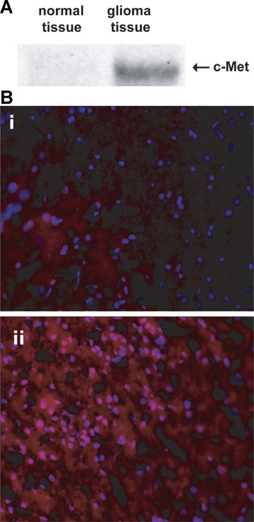
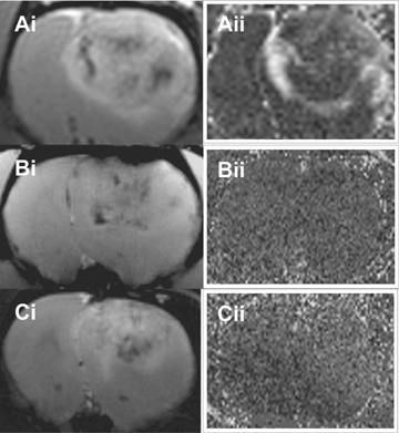
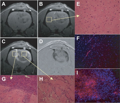
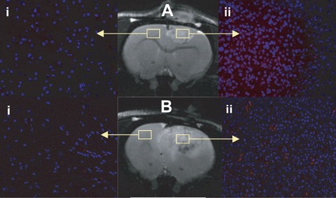
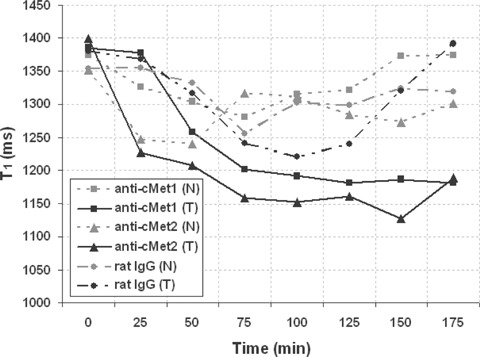
The tyrosine kinase receptor, c-Met, and its substrate, the hepatocyte growth factor (HGF), are implicated in the malignant progression of glioblastomas. In vivo detection of c-Met expression may be helpful in the diagnosis of malignant tumours. The C6 rat glioma model is a widely used intracranial brain tumour model used to study gliomas experimentally. We used a magnetic resonance imaging (MRI) molecular targeting agent to specifically tag the cell surface receptor, c-Met, with an anti-c-Met antibody (Ab) linked to biotinylated Gd (gadolinium)-DTPA (diethylene triamine penta acetic acid)-albumin in rat gliomas to detect overexpression of this antigen in vivo. The anti-c-Met probe (anti-c-Met-Gd-DTPA-albumin) was administered intravenously, and as determined by an increase in MRI signal intensity and a corresponding decrease in regional T(1) relaxation values, this probe was found to detect increased expression of c-Met protein levels in C6 gliomas. In addition, specificity for the binding of the anti-c-Met contrast agent was determined by using fluorescence microscopic imaging of the biotinylated portion of the targeting agent within neoplastic and 'normal'brain tissues following in vivo administration of the anti-c-Met probe. Controls with no Ab or with a normal rat IgG attached to the contrast agent component indicated no non-specific binding to glioma tissue. This is the first successful visualization of in vivo overexpression of c-Met in gliomas.
酪氨酸激酶受体c-Met及其底物肝细胞生长因子(HGF)与胶质母细胞瘤的恶性进展有关。体内检测c-Met表达可能有助于恶性肿瘤的诊断。C6大鼠胶质瘤模型是一种广泛用于实验性研究胶质瘤的颅内脑肿瘤模型。我们使用磁共振成像(MRI)分子靶向剂,通过将抗c-Met抗体(Ab)与生物素化的钆(Gd)-二乙三胺五乙酸(DTPA)-白蛋白相连,特异性标记大鼠胶质瘤细胞表面受体c-Met,以检测该抗原在体内的过表达。静脉注射抗c-Met探针(抗c-Met-Gd-DTPA-白蛋白),通过MRI信号强度增加和区域T(1)弛豫值相应降低确定,该探针可检测C6胶质瘤中c-Met蛋白水平的增加表达。此外,在体内注射抗c-Met探针后,通过对肿瘤和“正常”脑组织中靶向剂生物素化部分进行荧光显微镜成像,确定抗c-Met造影剂结合的特异性。未连接Ab或连接正常大鼠IgG的造影剂成分对照组显示与胶质瘤组织无非特异性结合。这是首次成功在体内可视化胶质瘤中c-Met的过表达。