Zhang Dongsheng, Fan Guo-Chang, Zhou Xiaoyang, Zhao Tiemin, Pasha Zeeshan, Xu Meifeng, Zhu Yi, Ashraf Muhammad, Wang Yigang
Department of Pathology and Laboratory Medicine, University of Cincinnati Medical Center, 231 Albert Sabin Way, Cincinnati, OH 45267-0529, USA.
J Mol Cell Cardiol. 2008 Feb;44(2):281-92. doi: 10.1016/j.yjmcc.2007.11.010. Epub 2007 Dec 7.
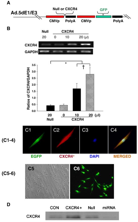
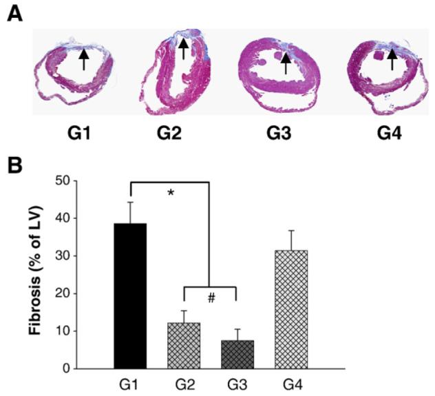
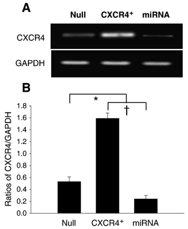
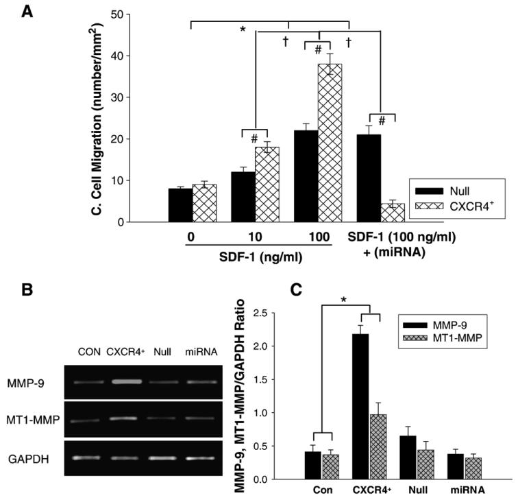
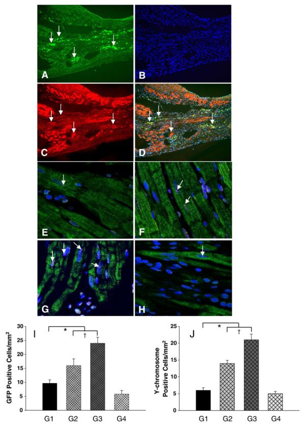
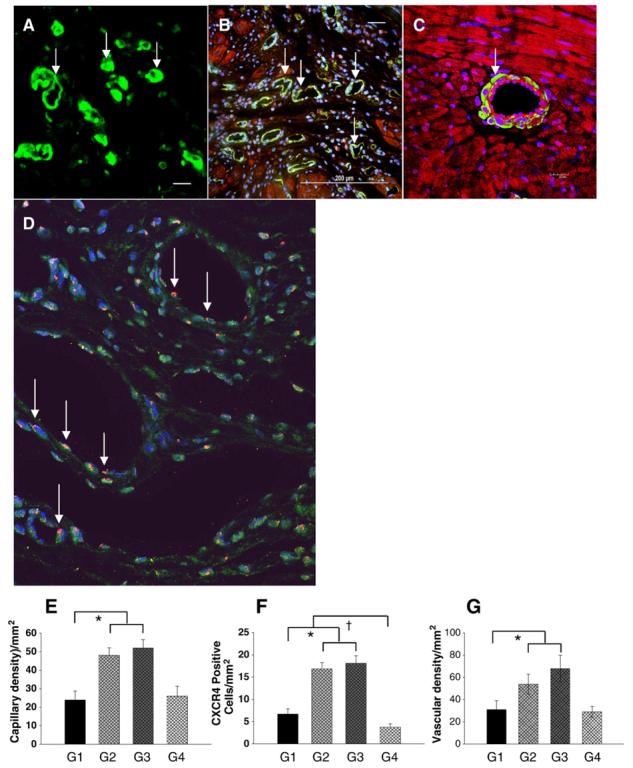
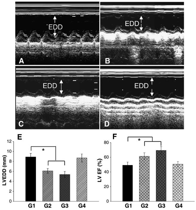
Bone marrow mesenchymal stem cells (MSCs) participate in myocardial repair following myocardial infarction. However, their in vivo reparative capability is limited due to lack of their survival in the infarcted myocardium. To overcome this limitation, we genetically engineered male rat MSCs overexpressing CXCR4 in order to maximize the effect of stromal cell-derived factor-1alpha (SDF-1alpha) for cell migration and regeneration. MSCs were isolated from adult male rats and cultured. Adenoviral transduction was carried out to over-express either CXCR4/green fluorescent protein (Ad-CXCR4/GFP) or Ad-null/GFP alone (control). Flow cytometry was used to identify and isolate GFP/CXCR4 over-expressing MSCs for transplantation. Female rats were assigned to one of four groups (n=8 each) to receive GFP-transduced male MSCs (2 x 10(6)) via tail vein injection 3 days after ligation of the left anterior descending (LAD) coronary artery: GFP-transduced MSCs (Ad-null/GFP-MSCs, group 1) or MSCs over-expressing CXCR4/GFP (Ad-CXCR4/GFP-MSCs, group 2), or Ad-CXCR4/GFP-MSCs plus SDF-1alpha (50 ng/microl) (Ad-CXCR4/GFP-MSCs/SDF-1alpha, group 3), or Ad-miRNA targeting CXCR4 plus SDF-1alpha (Ad-miRNA/GFP-MSCs+SDF-1alpha treatment, group 4). Cardiodynamic data were obtained 4 weeks after induction of regional myocardial infarction (MI) using echocardiography after which hearts were harvested for immunohistochemical studies. The migration of GFP and Y-chromosome positive cells increased significantly in the peri- and infarct areas of groups 2 and 3 compared to control group (p<0.05), or miRNA-CXCR4 group (p<0.01). The number of CXCR4 positive cells in groups 2, 3 was intimately associated with angiogenesis and myogenesis. MSCs engraftment was blocked by pretreatment with miRNA (group 4). Cardiac function was significantly improved in rats receiving MSCs over-expressing CXCR4 alone or with SDF-1alpha. The up-regulation of matrix metalloproteinases (MMPs) by CXCR4 overexpressing MSCs perhaps facilitated their engraftment in the collagenous tissue of the infarcted area. CXCR4 over-expression led to enhance in vivo mobilization and engraftment of MSCs into ischemic area where these cells promoted neomyoangiogenesis and alleviated early signs of left ventricular remodeling.
骨髓间充质干细胞(MSCs)参与心肌梗死后的心肌修复。然而,由于它们在梗死心肌中难以存活,其体内修复能力有限。为克服这一限制,我们对雄性大鼠MSCs进行基因工程改造,使其过表达CXCR4,以最大化基质细胞衍生因子-1α(SDF-1α)对细胞迁移和再生的作用。从成年雄性大鼠中分离并培养MSCs。进行腺病毒转导以单独过表达CXCR4/绿色荧光蛋白(Ad-CXCR4/GFP)或仅过表达Ad-空载体/GFP(对照)。使用流式细胞术鉴定并分离过表达GFP/CXCR4的MSCs用于移植。雌性大鼠被分为四组之一(每组n = 8),在左冠状动脉前降支(LAD)结扎3天后通过尾静脉注射接受GFP转导的雄性MSCs(2×10⁶):GFP转导的MSCs(Ad-空载体/GFP-MSCs,第1组)或过表达CXCR4/GFP的MSCs(Ad-CXCR4/GFP-MSCs,第2组),或Ad-CXCR4/GFP-MSCs加SDF-1α(50 ng/μl)(Ad-CXCR4/GFP-MSCs/SDF-1α,第3组),或靶向CXCR4的Ad-miRNA加SDF-1α(Ad-miRNA/GFP-MSCs + SDF-1α处理,第4组)。在诱导局部心肌梗死(MI)4周后,使用超声心动图获得心脏动力学数据,之后收获心脏进行免疫组织化学研究。与对照组(p < 0.05)或miRNA-CXCR4组(p < 0.01)相比,第2组和第3组梗死周边和梗死区域中GFP和Y染色体阳性细胞的迁移显著增加。第2组、第3组中CXCR4阳性细胞的数量与血管生成和肌生成密切相关。用miRNA预处理可阻断MSCs的植入(第4组)。单独接受过表达CXCR4的MSCs或与SDF-1α联合接受MSCs的大鼠心脏功能显著改善。过表达CXCR4的MSCs对基质金属蛋白酶(MMPs)的上调可能促进了它们在梗死区域胶原组织中的植入。CXCR4的过表达导致MSCs在体内向缺血区域的动员和植入增强,这些细胞在缺血区域促进了新生血管生成并减轻了左心室重构的早期迹象。