• 文献检索
  • 文档翻译
  • 深度研究
  • 学术资讯
  • Suppr Zotero 插件Zotero 插件
  • 邀请有礼
  • 套餐&价格
  • 历史记录
应用&插件
Suppr Zotero 插件Zotero 插件浏览器插件Mac 客户端Windows 客户端微信小程序
定价
高级版会员购买积分包购买API积分包
服务
文献检索文档翻译深度研究API 文档MCP 服务
关于我们
关于 Suppr公司介绍联系我们用户协议隐私条款
关注我们

Suppr 超能文献

核心技术专利:CN118964589B侵权必究
粤ICP备2023148730 号-1Suppr @ 2026

文献检索

告别复杂PubMed语法,用中文像聊天一样搜索,搜遍4000万医学文献。AI智能推荐,让科研检索更轻松。

立即免费搜索

文件翻译

保留排版,准确专业,支持PDF/Word/PPT等文件格式,支持 12+语言互译。

免费翻译文档

深度研究

AI帮你快速写综述,25分钟生成高质量综述,智能提取关键信息,辅助科研写作。

立即免费体验

自杀基因揭示了过表达 CXCR4 的间充质干细胞(MSC(CXCR4))的心肌血管新生作用。

Suicide gene reveals the myocardial neovascularization role of mesenchymal stem cells overexpressing CXCR4 (MSC(CXCR4)).

机构信息

Department of Pathology, College of Medicine, University of Cincinnati Medical Center, Cincinnati, Ohio, United States of America.

出版信息

PLoS One. 2012;7(9):e46158. doi: 10.1371/journal.pone.0046158. Epub 2012 Sep 28.

DOI:10.1371/journal.pone.0046158
PMID:23029422
原文链接:https://pmc.ncbi.nlm.nih.gov/articles/PMC3460871/
Abstract

BACKGROUND

Our previous studies indicated that MSC(CXCR4) improved cardiac function after myocardial infarction (MI). This study was aimed to investigate the specific role of MSC(CXCR4) in neovascularization of infarcted myocardium using a suicide gene approach.

METHODS

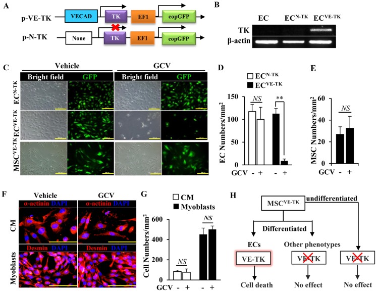
MSCs were transduced with either lentivirus-null vector/GFP (MSC(Null) as control) or vector encoding for overexpressing CXCR4/GFP. The MSC derived-endothelial cell (EC) differentiation was assessed by a tube formation assay, Dil-ac-LDL uptake, EC marker expression, and VE-cadherin promoter activity assay. Gene expression was analyzed by quantitative RT-PCR or Western blot. The suicide gene approach was under the control of VE-cadherin promoter. In vivo studies: Cell patches containing MSC(Null) or MSC(CXCR4) were transduced with suicide gene and implanted into the myocardium of MI rat. Rats received either ganciclovir (GCV) or vehicle after cell implantation. After one month, the cardiac functional changes and neovascularization were assessed by echocardiography, histological analysis, and micro-CT imaging.

RESULTS

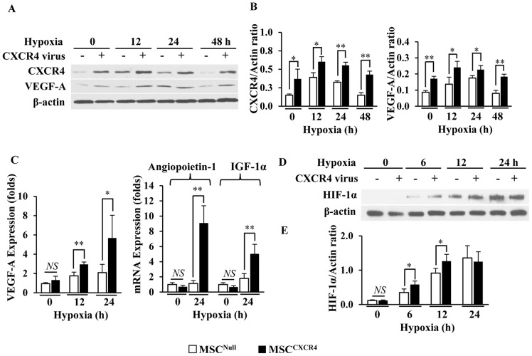
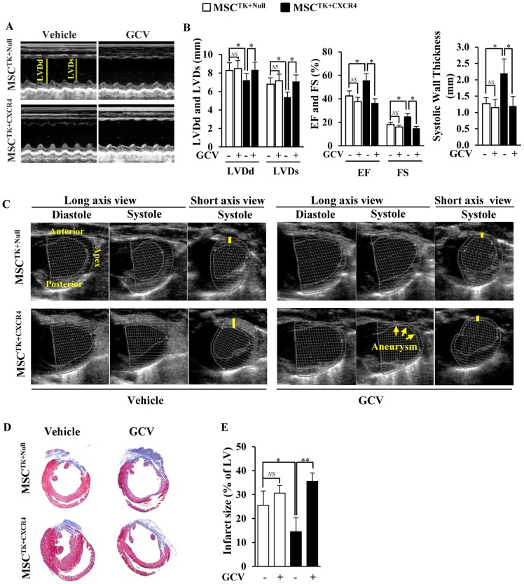
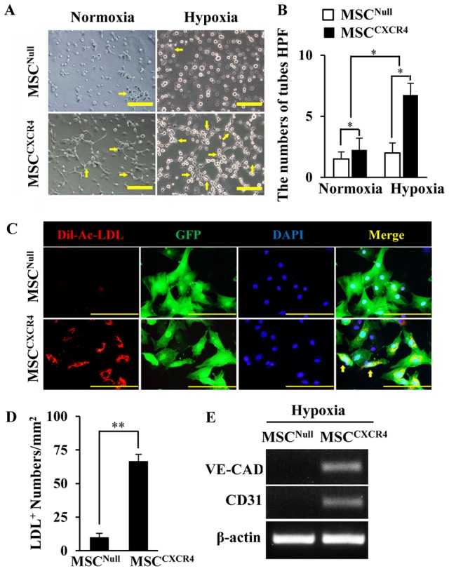
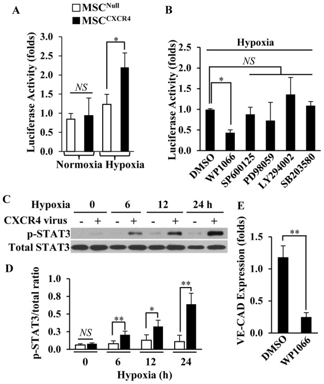
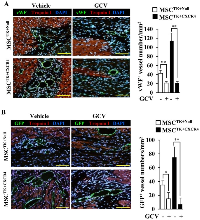
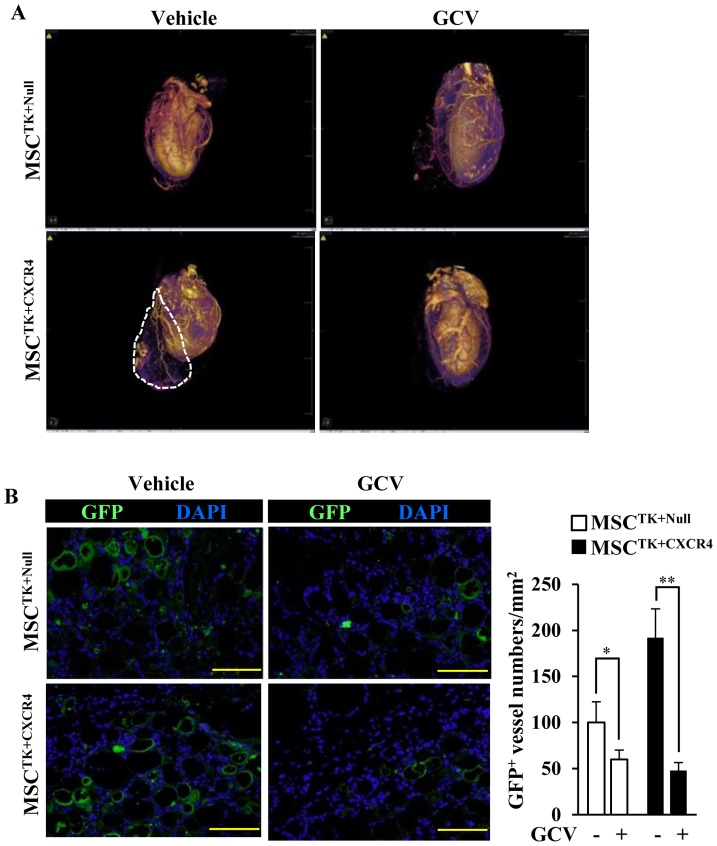
The expression of VEGF-A and HIF-1α was significantly higher in MSC(CXCR4) as compared to MSC(Null) under hypoxia. Additionally, MSC(CXCR4) enhanced new vessel formation and EC differentiation, as well as STAT3 phosphorylation under hypoxia. STAT3 participated in the transcription of VE-cadherin in MSC(CXCR4) under hypoxia, which was inhibited by WP1066 (a STAT3 inhibitor). In addition, GCV specifically induced death of ECs with suicide gene activation. In vivo studies: MSC(CXCR4) implantation promoted cardiac functional restoration, reduced infarct size, improved cardiac remodeling, and enhanced neovascularization in ischemic heart tissue. New vessels derived from MSC(CXCR4) were observed at the injured heart margins and communicated with native coronary arteries. However, the derived vessel networks were reduced by GCV, reversing improvement of cardiac function.

CONCLUSION

The transplanted MSC(CXCR4) enhanced neovascularization after MI by boosting release of angiogenic factors and increasing the potential of endothelial differentiation.

摘要

背景

我们之前的研究表明 MSC(CXCR4)可改善心肌梗死(MI)后的心脏功能。本研究旨在通过自杀基因方法探讨 MSC(CXCR4)在梗死心肌新生血管形成中的具体作用。

方法

通过慢病毒空载体/GFP(MSC(Null)作为对照)或载体转染过表达 CXCR4/GFP 的 MSC。通过管形成试验、Dil-ac-LDL 摄取、内皮细胞标志物表达和 VE-cadherin 启动子活性测定评估 MSC 衍生的内皮细胞(EC)分化。通过定量 RT-PCR 或 Western blot 分析基因表达。自杀基因方法受 VE-cadherin 启动子控制。体内研究:将含有 MSC(Null)或 MSC(CXCR4)的细胞片用自杀基因转导后植入 MI 大鼠的心肌中。细胞植入后,大鼠接受更昔洛韦(GCV)或载体。一个月后,通过超声心动图、组织学分析和 micro-CT 成像评估心脏功能变化和新生血管形成。

结果

与 MSC(Null)相比,缺氧时 MSC(CXCR4)中 VEGF-A 和 HIF-1α 的表达明显更高。此外,缺氧下 MSC(CXCR4)增强了新血管形成和 EC 分化,以及 STAT3 磷酸化。STAT3 参与了缺氧下 MSC(CXCR4)中 VE-cadherin 的转录,WP1066(STAT3 抑制剂)抑制了这一转录。此外,GCV 可通过自杀基因激活特异性诱导 EC 死亡。体内研究:MSC(CXCR4)植入可促进心脏功能恢复、减少梗死面积、改善心脏重构,并增强缺血心肌组织中的新生血管形成。在受伤的心脏边缘观察到源自 MSC(CXCR4)的新血管,并与原生冠状动脉相通。然而,GCV 减少了衍生的血管网络,逆转了心脏功能的改善。

结论

移植的 MSC(CXCR4)通过增加血管生成因子的释放和增加内皮分化的潜力,增强 MI 后的新生血管形成。

https://cdn.ncbi.nlm.nih.gov/pmc/blobs/cf21/3460871/1e37f96f8b3a/pone.0046158.g007.jpg
https://cdn.ncbi.nlm.nih.gov/pmc/blobs/cf21/3460871/701593d1e7eb/pone.0046158.g001.jpg
https://cdn.ncbi.nlm.nih.gov/pmc/blobs/cf21/3460871/860732b93d95/pone.0046158.g002.jpg
https://cdn.ncbi.nlm.nih.gov/pmc/blobs/cf21/3460871/b223d1e0e6e2/pone.0046158.g003.jpg
https://cdn.ncbi.nlm.nih.gov/pmc/blobs/cf21/3460871/d9652e72208f/pone.0046158.g004.jpg
https://cdn.ncbi.nlm.nih.gov/pmc/blobs/cf21/3460871/bfddb762ada1/pone.0046158.g005.jpg
https://cdn.ncbi.nlm.nih.gov/pmc/blobs/cf21/3460871/043065329328/pone.0046158.g006.jpg
https://cdn.ncbi.nlm.nih.gov/pmc/blobs/cf21/3460871/1e37f96f8b3a/pone.0046158.g007.jpg

相似文献

1
Suicide gene reveals the myocardial neovascularization role of mesenchymal stem cells overexpressing CXCR4 (MSC(CXCR4)).自杀基因揭示了过表达 CXCR4 的间充质干细胞(MSC(CXCR4))的心肌血管新生作用。
PLoS One. 2012;7(9):e46158. doi: 10.1371/journal.pone.0046158. Epub 2012 Sep 28.
2
Paracrine effect of CXCR4-overexpressing mesenchymal stem cells on ischemic heart injury.CXCR4过表达间充质干细胞对缺血性心脏损伤的旁分泌作用。
Cell Biochem Funct. 2017 Mar;35(2):113-123. doi: 10.1002/cbf.3254. Epub 2017 Feb 23.
3
Over-expression of CXCR4 on mesenchymal stem cells augments myoangiogenesis in the infarcted myocardium.间充质干细胞上CXCR4的过表达增强梗死心肌中的肌血管生成。
J Mol Cell Cardiol. 2008 Feb;44(2):281-92. doi: 10.1016/j.yjmcc.2007.11.010. Epub 2007 Dec 7.
4
Genetically manipulated progenitor cell sheet with diprotin A improves myocardial function and repair of infarcted hearts.携带二肽基肽酶 A 的基因修饰祖细胞片改善心肌功能和梗死心脏修复。
Am J Physiol Heart Circ Physiol. 2010 Nov;299(5):H1339-47. doi: 10.1152/ajpheart.00592.2010. Epub 2010 Aug 27.
5
Mesenchymal stem cells overexpressing CXCR4 attenuate remodeling of postmyocardial infarction by releasing matrix metalloproteinase-9.过表达 CXCR4 的间充质干细胞通过释放基质金属蛋白酶-9 减轻心肌梗死后的重构。
Stem Cells Dev. 2012 Mar 20;21(5):778-89. doi: 10.1089/scd.2011.0126. Epub 2011 Jul 22.
6
Thioredoxin-1 (Trx1) engineered mesenchymal stem cell therapy increased pro-angiogenic factors, reduced fibrosis and improved heart function in the infarcted rat myocardium.硫氧还蛋白-1(Trx1)工程化间充质干细胞疗法增加了促血管生成因子,减少了纤维化,并改善了梗死大鼠心肌的心脏功能。
Int J Cardiol. 2015 Dec 15;201:517-28. doi: 10.1016/j.ijcard.2015.08.117. Epub 2015 Aug 15.
7
VEGF/SDF-1 promotes cardiac stem cell mobilization and myocardial repair in the infarcted heart.VEGF/SDF-1 促进心脏干细胞动员和梗死心脏修复。
Cardiovasc Res. 2011 Aug 1;91(3):402-11. doi: 10.1093/cvr/cvr053. Epub 2011 Feb 22.
8
MicroRNA-377 regulates mesenchymal stem cell-induced angiogenesis in ischemic hearts by targeting VEGF.微小RNA-377通过靶向血管内皮生长因子调控缺血心脏中间充质干细胞诱导的血管生成。
PLoS One. 2014 Sep 24;9(9):e104666. doi: 10.1371/journal.pone.0104666. eCollection 2014.
9
Paracrine factors released by GATA-4 overexpressed mesenchymal stem cells increase angiogenesis and cell survival.过表达 GATA-4 的间充质干细胞释放的旁分泌因子可增加血管生成和细胞存活。
Am J Physiol Heart Circ Physiol. 2010 Dec;299(6):H1772-81. doi: 10.1152/ajpheart.00557.2010. Epub 2010 Sep 24.
10
Myocardial transfection of hypoxia-inducible factor-1α and co-transplantation of mesenchymal stem cells enhance cardiac repair in rats with experimental myocardial infarction.缺氧诱导因子-1α的心肌转染及间充质干细胞的共移植增强实验性心肌梗死大鼠的心脏修复
Stem Cell Res Ther. 2014 Feb 7;5(1):22. doi: 10.1186/scrt410.

引用本文的文献

1
Therapeutic Applications of Engineered Mesenchymal Stromal Cells for Enhanced Angiogenesis in Cardiac and Cerebral Ischemia.工程化间充质基质细胞在心脏和脑缺血中增强血管生成的治疗应用。
Stem Cell Rev Rep. 2024 Nov;20(8):2138-2154. doi: 10.1007/s12015-024-10787-3. Epub 2024 Sep 21.
2
Nanoparticles and Mesenchymal Stem Cell (MSC) Therapy for Cancer Treatment: Focus on Nanocarriers and a si-RNA CXCR4 Chemokine Blocker as Strategies for Tumor Eradication In Vitro and In Vivo.纳米颗粒与间充质干细胞(MSC)疗法用于癌症治疗:聚焦纳米载体以及作为体内外根除肿瘤策略的si-RNA CXCR4趋化因子阻断剂
Micromachines (Basel). 2023 Nov 7;14(11):2068. doi: 10.3390/mi14112068.
3

本文引用的文献

1
Induction of vascular progenitor cells from endothelial cells stimulates coronary collateral growth.从内皮细胞诱导血管祖细胞可刺激冠状动脉侧支生长。
Circ Res. 2012 Jan 20;110(2):241-52. doi: 10.1161/CIRCRESAHA.111.250126. Epub 2011 Nov 17.
2
Reduced collagen deposition in infarcted myocardium facilitates induced pluripotent stem cell engraftment and angiomyogenesis for improvement of left ventricular function.梗死心肌中胶原蛋白沉积减少有助于诱导多能干细胞的植入和血管生成,从而改善左心室功能。
J Am Coll Cardiol. 2011 Nov 8;58(20):2118-27. doi: 10.1016/j.jacc.2011.06.062.
3
Molecular control of endothelial cell behaviour during blood vessel morphogenesis.
Modifying strategies for SDF-1/CXCR4 interaction during mesenchymal stem cell transplantation.
修改间充质干细胞移植过程中 SDF-1/CXCR4 相互作用的策略。
Gen Thorac Cardiovasc Surg. 2022 Jan;70(1):1-10. doi: 10.1007/s11748-021-01696-0. Epub 2021 Sep 12.
4
Mapping current research and identifying hotspots on mesenchymal stem cells in cardiovascular disease.绘制当前研究图谱并确定心血管疾病间充质干细胞的热点。
Stem Cell Res Ther. 2020 Nov 25;11(1):498. doi: 10.1186/s13287-020-02009-7.
5
Genetic Engineering as a Strategy to Improve the Therapeutic Efficacy of Mesenchymal Stem/Stromal Cells in Regenerative Medicine.基因工程作为提高间充质干/基质细胞在再生医学中治疗效果的一种策略。
Front Cell Dev Biol. 2020 Aug 21;8:737. doi: 10.3389/fcell.2020.00737. eCollection 2020.
6
MicroRNA-133 overexpression promotes the therapeutic efficacy of mesenchymal stem cells on acute myocardial infarction.MicroRNA-133 过表达促进间充质干细胞对急性心肌梗死的治疗效果。
Stem Cell Res Ther. 2017 Nov 25;8(1):268. doi: 10.1186/s13287-017-0722-z.
7
Effects of miR-146a on the osteogenesis of adipose-derived mesenchymal stem cells and bone regeneration.miR-146a 对脂肪间充质干细胞成骨分化及骨再生的影响。
Sci Rep. 2017 Feb 16;7:42840. doi: 10.1038/srep42840.
8
Electrical Stimulation Enhances Cardiac Differentiation of Human Induced Pluripotent Stem Cells for Myocardial Infarction Therapy.电刺激增强人诱导多能干细胞向心肌梗死治疗中的心肌分化。
Antioxid Redox Signal. 2018 Feb 10;28(5):371-384. doi: 10.1089/ars.2016.6766. Epub 2017 Jan 12.
9
Mesenchymal stem cells in cardiac regeneration: a detailed progress report of the last 6 years (2010-2015).心脏再生中的间充质干细胞:过去6年(2010 - 2015年)的详细进展报告
Stem Cell Res Ther. 2016 Jun 4;7(1):82. doi: 10.1186/s13287-016-0341-0.
10
Implantation of a Novel Allogeneic Mesenchymal Precursor Cell Type in Patients with Ischemic Cardiomyopathy Undergoing Coronary Artery Bypass Grafting: an Open Label Phase IIa Trial.新型同种异体间充质前体细胞类型植入接受冠状动脉旁路移植术的缺血性心肌病患者:一项开放标签IIa期试验。
J Cardiovasc Transl Res. 2016 Jun;9(3):202-213. doi: 10.1007/s12265-016-9686-0. Epub 2016 Apr 1.
血管生成过程中内皮细胞行为的分子调控。
Nat Rev Mol Cell Biol. 2011 Aug 23;12(9):551-64. doi: 10.1038/nrm3176.
4
SDF-1/CXCR4 mediates acute protection of cardiac function through myocardial STAT3 signaling following global ischemia/reperfusion injury.基质细胞衍生因子 1/CXCR4 通过心肌 STAT3 信号转导介导缺血/再灌注损伤后心脏功能的急性保护。
Am J Physiol Heart Circ Physiol. 2011 Oct;301(4):H1496-505. doi: 10.1152/ajpheart.00365.2011. Epub 2011 Aug 5.
5
The MSC: an injury drugstore.MSC:损伤药物库。
Cell Stem Cell. 2011 Jul 8;9(1):11-5. doi: 10.1016/j.stem.2011.06.008.
6
STAT3/Pim-1 signaling pathway plays a crucial role in endothelial differentiation of cardiac resident Sca-1+ cells both in vitro and in vivo.STAT3/Pim-1 信号通路在心脏驻留 Sca-1+ 细胞的内皮分化中具有重要作用,无论是在体外还是体内。
J Mol Cell Cardiol. 2011 Aug;51(2):207-14. doi: 10.1016/j.yjmcc.2011.04.013. Epub 2011 May 6.
7
The elusive nature and function of mesenchymal stem cells.间充质干细胞的隐匿性质和功能。
Nat Rev Mol Cell Biol. 2011 Feb;12(2):126-31. doi: 10.1038/nrm3049.
8
Hypoxia-inducible factors and the response to hypoxic stress.缺氧诱导因子与应对缺氧应激。
Mol Cell. 2010 Oct 22;40(2):294-309. doi: 10.1016/j.molcel.2010.09.022.
9
Overexpression of phosphoinositide-3-kinase class II alpha enhances mesenchymal stem cell survival in infarcted myocardium.磷酸肌醇-3-激酶 II 亚型的过表达增强骨髓间充质干细胞在梗死心肌中的存活。
Biochem Biophys Res Commun. 2010 Nov 12;402(2):272-9. doi: 10.1016/j.bbrc.2010.10.013. Epub 2010 Oct 16.
10
Activation of host tissue trophic factors through JAK-STAT3 signaling: a mechanism of mesenchymal stem cell-mediated cardiac repair.通过 JAK-STAT3 信号转导激活宿主组织营养因子:间充质干细胞介导的心脏修复机制。
Am J Physiol Heart Circ Physiol. 2010 Nov;299(5):H1428-38. doi: 10.1152/ajpheart.00488.2010. Epub 2010 Sep 17.