Hostmann Arwed, Jasse Kerstin, Schulze-Tanzil Gundula, Robinson Yohan, Oberholzer Andreas, Ertel Wolfgang, Tschoeke Sven K
Department of Trauma and Reconstructive Surgery, Charité-University Medical School Berlin, Campus Benjamin Franklin, 12207 Berlin, Germany.
Crit Care. 2008;12(1):R8. doi: 10.1186/cc6772. Epub 2008 Jan 22.
The innate immune response to trauma hemorrhage involves inflammatory mediators, thus promoting cellular dysfunction as well as cell death in diverse tissues. These effects ultimately bear the risk of post-traumatic complications such as organ dysfunction, multiple organ failure, or adult respiratory distress syndrome. In this study, a murine model of resuscitated hemorrhagic shock (HS) was used to determine the apoptosis in spleen as a marker of cellular injury and reduced immune functions.
Male C57BL-6 mice were subjected to sham operation or resuscitated HS. At t = 0 hours, t = 24 hours, and t = 72 hours, mice were euthanized and the spleens were removed and evaluated for apoptotic changes via DNA fragmentation, caspase activities, and activation of both extrinsic and intrinsic apoptotic pathways. Spleens from untreated mice were used as control samples.
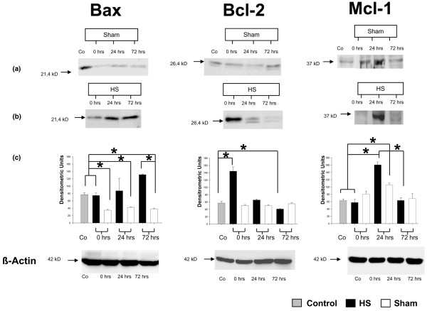
HS was associated with distinct lymphocytopenia as early as t = 0 hours after hemorrhage without regaining baseline levels within the consecutive 72 hours when compared with sham and control groups. A rapid activation of splenic apoptosis in HS mice was observed at t = 0 hours and t = 72 hours after hemorrhage and predominantly confirmed by increased DNA fragmentation, elevated caspase-3/7, caspase-8, and caspase-9 activities, and enhanced expression of intrinsic mitochondrial proteins. Accordingly, mitochondrial pro-apoptotic Bax and anti-apoptotic Bcl-2 proteins were inversely expressed within the 72-hour observation period, thereby supporting significant pro-apoptotic changes. Solely at t = 24 hours, expression of the anti-apoptotic Mcl-1 protein shows a significant increase when compared with sham-operated and control animals. Furthermore, expression of extrinsic death receptors were only slightly increased.
Our data suggest that HS induces apoptotic changes in spleen through a biphasic caspase-dependent mechanism and imply a detrimental imbalance of pro- and anti-apoptotic mitochondrial proteins Bax, Bcl-2, and Mcl-1, thereby promoting post-traumatic immunosuppression.
机体对创伤性出血的固有免疫反应涉及炎症介质,从而促进多种组织中的细胞功能障碍以及细胞死亡。这些效应最终会带来创伤后并发症的风险,如器官功能障碍、多器官功能衰竭或成人呼吸窘迫综合征。在本研究中,使用复苏性失血性休克(HS)小鼠模型来确定脾脏中的细胞凋亡情况,以此作为细胞损伤和免疫功能降低的标志物。
对雄性C57BL-6小鼠进行假手术或复苏性HS处理。在t = 0小时、t = 24小时和t = 72小时时,对小鼠实施安乐死并取出脾脏,通过DNA片段化、半胱天冬酶活性以及外源性和内源性凋亡途径的激活来评估凋亡变化。将未处理小鼠的脾脏用作对照样本。
与假手术组和对照组相比,HS早在出血后t = 0小时就伴有明显的淋巴细胞减少,且在随后的72小时内未恢复到基线水平。在出血后t = 0小时和t = 72小时观察到HS小鼠脾脏凋亡迅速激活,主要表现为DNA片段化增加、半胱天冬酶-3/7、半胱天冬酶-8和半胱天冬酶-9活性升高以及内源性线粒体蛋白表达增强。相应地,线粒体促凋亡蛋白Bax和抗凋亡蛋白Bcl-2在72小时观察期内呈反向表达,从而支持了显著的促凋亡变化。仅在t = 24小时时,与假手术组和对照动物相比,抗凋亡蛋白Mcl-1的表达显著增加。此外,外源性死亡受体的表达仅略有增加。
我们的数据表明,HS通过双相半胱天冬酶依赖性机制诱导脾脏凋亡变化,并提示促凋亡和抗凋亡线粒体蛋白Bax、Bcl-2和Mcl-1存在有害失衡,从而促进创伤后免疫抑制。