Department of Radiology, Massachusetts General Hospital and Harvard Medical School, White 270 55 Fruit Street, Boston, MA 02114, USA.
Cancer Imaging. 2004 Apr 2;4 Spec No A(Spec No A):S42-6. doi: 10.1102/1470-7330.2004.0011.
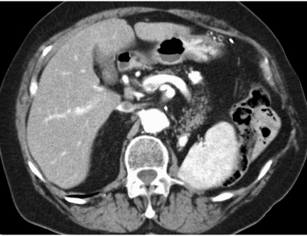
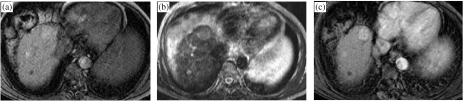
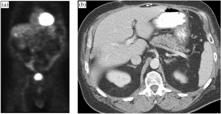
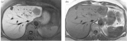
Liver imaging in patients with a history of known or suspected malignancy is important because the liver is a common site of metastatic spread, especially tumours from the colon, lung, pancreas and stomach, and in patients with chronic liver disease who are at risk for developing hepatocellular carcinoma. Since benign liver lesions are common, liverimaging strategies should incorporate liver lesion detection and characterisation. Survey examination in patients with a known extra-hepatic malignancy to exclude the presence of hepatic and extra-hepatic involvement is normally undertaken with a contrast-enhanced computed tomography examination. When patients with hepatic metastases are being considered for metastasesectomy, they undergo a staging examination with contrast-enhanced magnetic resonance imaging (MRI) using tissue-specific contrast agents. Patients with chronic liver disease who are at risk for hepatocellular carcinoma undergo periodic liver screening for focal liver detection, usually with ultrasonography (US) with MRI being used when US is equivocal. Finally, contrast-enhanced MRI with extra-cellular gadolinium chelates is preferred for characterisation of indeterminate hepatic masses with liver biopsy used when tissue diagnosis is needed.
对于有已知或疑似恶性肿瘤病史的患者,肝脏影像学检查很重要,因为肝脏是转移扩散的常见部位,特别是来自结肠、肺、胰腺和胃的肿瘤,以及患有慢性肝病、有发生肝细胞癌风险的患者。由于良性肝脏病变很常见,因此肝脏影像学检查策略应包括肝脏病变的检测和特征描述。对于有已知肝外恶性肿瘤的患者,通常采用增强 CT 检查进行全身检查,以排除肝内和肝外受累的情况。对于考虑进行肝转移瘤切除术的患者,会进行增强 MRI 检查,使用组织特异性对比剂进行分期检查。对于有发生肝细胞癌风险的慢性肝病患者,会进行定期的肝脏筛查,以检测局灶性肝脏病变,通常使用超声检查(US),当 US 结果不确定时,则使用 MRI。最后,对于不确定的肝脏肿块,使用细胞外钆螯合物增强 MRI 进行特征描述,当需要组织诊断时,则使用肝活检。