Macedo-Ribeiro Sandra, Almeida Carla, Calisto Bárbara M, Friedrich Thomas, Mentele Reinhard, Stürzebecher Jörg, Fuentes-Prior Pablo, Pereira Pedro José Barbosa
Centro de Neurociências e Biologia Celular (CNC), Coimbra, Portugal.
PLoS One. 2008 Feb 20;3(2):e1624. doi: 10.1371/journal.pone.0001624.
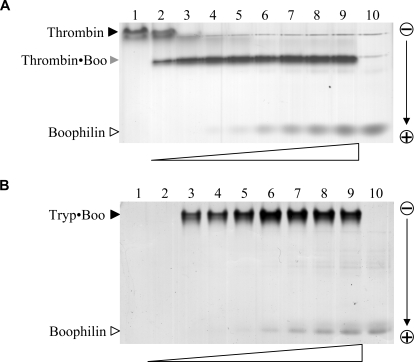
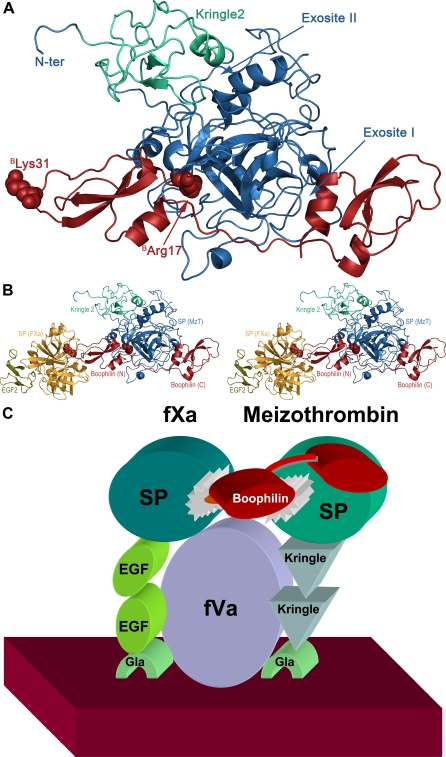
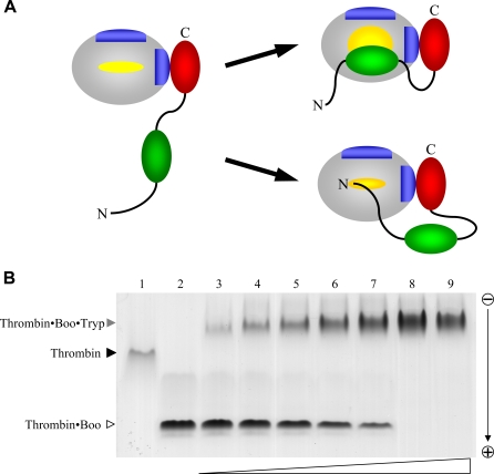
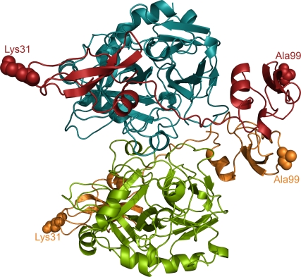
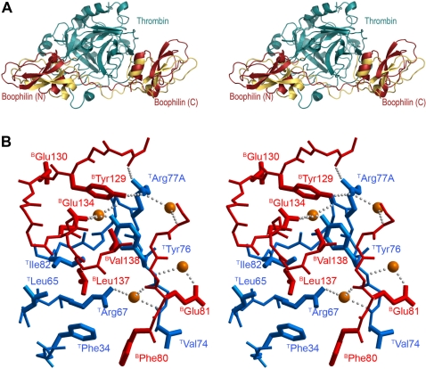
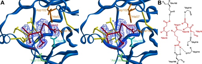
Inhibitors of coagulation factors from blood-feeding animals display a wide variety of structural motifs and inhibition mechanisms. We have isolated a novel inhibitor from the cattle tick Boophilus microplus, one of the most widespread parasites of farm animals. The inhibitor, which we have termed boophilin, has been cloned and overexpressed in Escherichia coli. Mature boophilin is composed of two canonical Kunitz-type domains, and inhibits not only the major procoagulant enzyme, thrombin, but in addition, and by contrast to all other previously characterised natural thrombin inhibitors, significantly interferes with the proteolytic activity of other serine proteinases such as trypsin and plasmin. The crystal structure of the bovine alpha-thrombin.boophilin complex, refined at 2.35 A resolution reveals a non-canonical binding mode to the proteinase. The N-terminal region of the mature inhibitor, Q16-R17-N18, binds in a parallel manner across the active site of the proteinase, with the guanidinium group of R17 anchored in the S(1) pocket, while the C-terminal Kunitz domain is negatively charged and docks into the basic exosite I of thrombin. This binding mode resembles the previously characterised thrombin inhibitor, ornithodorin which, unlike boophilin, is composed of two distorted Kunitz modules. Unexpectedly, both boophilin domains adopt markedly different orientations when compared to those of ornithodorin, in its complex with thrombin. The N-terminal boophilin domain rotates 9 degrees and is displaced by 6 A, while the C-terminal domain rotates almost 6 degrees accompanied by a 3 A displacement. The reactive-site loop of the N-terminal Kunitz domain of boophilin with its P(1) residue, K31, is fully solvent exposed and could thus bind a second trypsin-like proteinase without sterical restraints. This finding explains the formation of a ternary thrombin.boophilin.trypsin complex, and suggests a mechanism for prothrombinase inhibition in vivo.
来自吸血动物的凝血因子抑制剂呈现出各种各样的结构基序和抑制机制。我们从微小牛蜱(一种最为常见的家畜寄生虫)中分离出一种新型抑制剂。我们将这种抑制剂命名为牛蜱抗凝蛋白,它已在大肠杆菌中克隆并过量表达。成熟的牛蜱抗凝蛋白由两个典型的Kunitz型结构域组成,它不仅能抑制主要的促凝酶——凝血酶,而且与所有其他先前已鉴定的天然凝血酶抑制剂不同的是,它还能显著干扰其他丝氨酸蛋白酶(如胰蛋白酶和纤溶酶)的蛋白水解活性。牛α-凝血酶与牛蜱抗凝蛋白复合物的晶体结构,在2.35 Å分辨率下进行精修,揭示了其与蛋白酶的一种非典型结合模式。成熟抑制剂的N端区域,即Q16-R17-N18,以平行方式横跨蛋白酶的活性位点结合,R17的胍基锚定在S(1)口袋中,而C端Kunitz结构域带负电荷,对接至凝血酶的碱性外位点I。这种结合模式类似于先前鉴定的凝血酶抑制剂——Ornithodorin,不同的是,Ornithodorin由两个扭曲的Kunitz模块组成。出乎意料的是,与凝血酶形成复合物时,相比于Ornithodorin,牛蜱抗凝蛋白的两个结构域呈现出明显不同的取向。牛蜱抗凝蛋白的N端结构域旋转9度并位移6 Å,而C端结构域旋转近6度并伴有3 Å的位移。牛蜱抗凝蛋白N端Kunitz结构域的反应位点环及其P(1)残基K31完全暴露于溶剂中,因此可以在没有空间位阻的情况下结合第二个类胰蛋白酶。这一发现解释了三元凝血酶-牛蜱抗凝蛋白-胰蛋白酶复合物的形成,并提示了体内凝血酶原酶抑制的一种机制。