Risueño Ruth M, Schamel Wolfgang W A, Alarcón Balbino
Centro de Biología Molecular Severo Ochoa, Consejo Superior de Investigaciones Científicas, Universidad Autónoma de Madrid, Madrid, Spain.
PLoS One. 2008 Mar 5;3(3):e1747. doi: 10.1371/journal.pone.0001747.
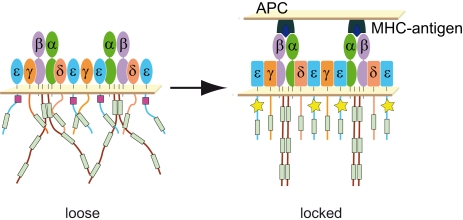
How the T cell antigen receptor (TCR) discriminates between molecularly related peptide/Major Histocompatibility Complex (pMHC) ligands and converts this information into different possible signaling outcomes is still not understood. One current model proposes that strong pMHC ligands, but not weak ones, induce a conformational change in the TCR. Evidence supporting this comes from a pull-down assay that detects ligand-induced binding of the TCR to the N-terminal SH3 domain of the adapter protein Nck, and also from studies with a neoepitope-specific antibody. Both methods rely on the exposure of a polyproline sequence in the CD3epsilon subunit of the TCR, and neither indicates whether the conformational change is transmitted to other CD3 subunits. Using a protease-sensitivity assay, we now show that the cytoplasmic tails of CD3epsilon and CD3zeta subunits become fully protected from degradation upon TCR triggering. These results suggest that the TCR conformational change is transmitted to the tails of CD3epsilon and CD3zeta, and perhaps all CD3 subunits. Furthermore, the resistance to protease digestion suggests that CD3 cytoplasmic tails adopt a compact structure in the triggered TCR. These results are consistent with a model in which transduction of the conformational change induced upon TCR triggering promotes condensation and shielding of the CD3 cytoplasmic tails.
T细胞抗原受体(TCR)如何区分分子相关的肽/主要组织相容性复合体(pMHC)配体,并将此信息转化为不同的可能信号转导结果,目前仍不清楚。当前一种模型提出,强pMHC配体而非弱配体可诱导TCR发生构象变化。支持这一观点的证据来自一项下拉实验,该实验检测到配体诱导TCR与衔接蛋白Nck的N端SH3结构域结合,也来自一项针对新表位特异性抗体的研究。这两种方法都依赖于TCR的CD3ε亚基中多聚脯氨酸序列的暴露,且均未表明构象变化是否传递至其他CD3亚基。通过蛋白酶敏感性实验,我们现在表明,TCR触发后,CD3ε和CD3ζ亚基的胞质尾完全受到保护不被降解。这些结果表明,TCR构象变化传递至CD3ε和CD3ζ的尾部,或许还有所有CD3亚基。此外,对蛋白酶消化的抗性表明,CD3胞质尾在被触发的TCR中采用紧凑结构。这些结果与一种模型一致,即TCR触发时诱导的构象变化的转导促进了CD3胞质尾的凝聚和屏蔽。