Ikonomovic Milos D, Klunk William E, Abrahamson Eric E, Mathis Chester A, Price Julie C, Tsopelas Nicholas D, Lopresti Brian J, Ziolko Scott, Bi Wenzhu, Paljug William R, Debnath Manik L, Hope Caroline E, Isanski Barbara A, Hamilton Ronald L, DeKosky Steven T
Department of Neurology, University of Pittsburgh School of Medicine, Pittsburgh, PA 15213, USA.
Brain. 2008 Jun;131(Pt 6):1630-45. doi: 10.1093/brain/awn016. Epub 2008 Mar 12.
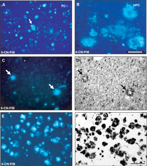
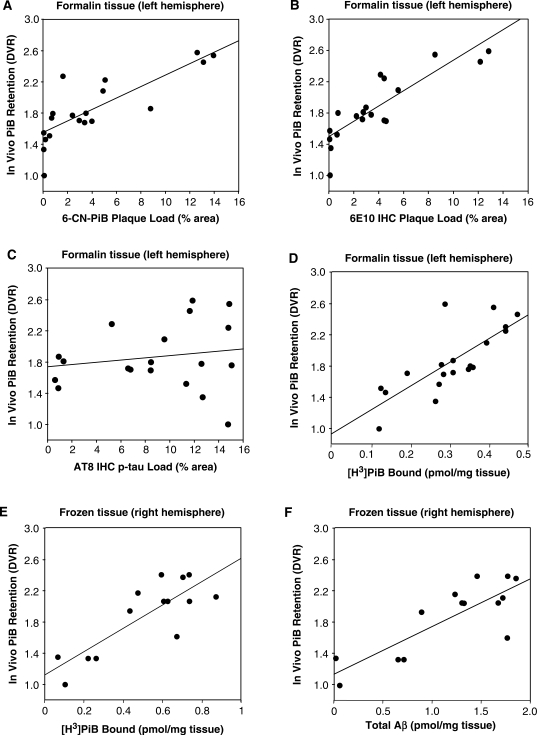
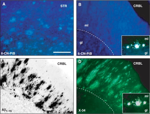
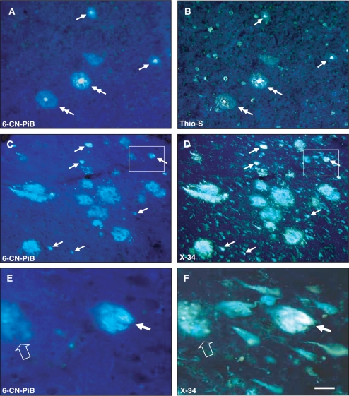
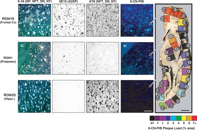
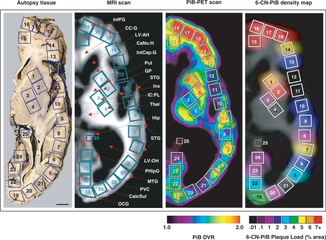
The positron emission tomography (PET) radiotracer Pittsburgh Compound-B (PiB) binds with high affinity to beta-pleated sheet aggregates of the amyloid-beta (Abeta) peptide in vitro. The in vivo retention of PiB in brains of people with Alzheimer's disease shows a regional distribution that is very similar to distribution of Abeta deposits observed post-mortem. However, the basis for regional variations in PiB binding in vivo, and the extent to which it binds to different types of Abeta-containing plaques and tau-containing neurofibrillary tangles (NFT), has not been thoroughly investigated. The present study examined 28 clinically diagnosed and autopsy-confirmed Alzheimer's disease subjects, including one Alzheimer's disease subject who had undergone PiB-PET imaging 10 months prior to death, to evaluate region- and substrate-specific binding of the highly fluorescent PiB derivative 6-CN-PiB. These data were then correlated with region-matched Abeta plaque load and peptide levels, [(3)H]PiB binding in vitro, and in vivo PET retention levels. We found that in Alzheimer's disease brain tissue sections, the preponderance of 6-CN-PiB binding is in plaques immunoreactive to either Abeta42 or Abeta40, and to vascular Abeta deposits. 6-CN-PiB labelling was most robust in compact/cored plaques in the prefrontal and temporal cortices. While diffuse plaques, including those in caudate nucleus and presubiculum, were less prominently labelled, amorphous Abeta plaques in the cerebellum were not detectable with 6-CN-PiB. Only a small subset of NFT were 6-CN-PiB positive; these resembled extracellular 'ghost' NFT. In Alzheimer's disease brain tissue homogenates, there was a direct correlation between [(3)H]PiB binding and insoluble Abeta peptide levels. In the Alzheimer's disease subject who underwent PiB-PET prior to death, in vivo PiB retention levels correlated directly with region-matched post-mortem measures of [(3)H]PiB binding, insoluble Abeta peptide levels, 6-CN-PiB- and Abeta plaque load, but not with measures of NFT. These results demonstrate, in a typical Alzheimer's disease brain, that PiB binding is highly selective for insoluble (fibrillar) Abeta deposits, and not for neurofibrillary pathology. The strong direct correlation of in vivo PiB retention with region-matched quantitative analyses of Abeta plaques in the same subject supports the validity of PiB-PET imaging as a method for in vivo evaluation of Abeta plaque burden.
正电子发射断层扫描(PET)放射性示踪剂匹兹堡化合物-B(PiB)在体外与β-淀粉样蛋白(Aβ)肽的β-折叠片聚集体具有高亲和力结合。PiB在阿尔茨海默病患者大脑中的体内滞留显示出一种区域分布,与死后观察到的Aβ沉积物分布非常相似。然而,PiB体内结合区域差异的基础,以及它与不同类型含Aβ斑块和含tau神经原纤维缠结(NFT)结合的程度,尚未得到充分研究。本研究检查了28名临床诊断并经尸检证实的阿尔茨海默病患者,包括一名在死亡前10个月接受过PiB-PET成像的阿尔茨海默病患者,以评估高荧光PiB衍生物6-CN-PiB的区域和底物特异性结合。然后将这些数据与区域匹配的Aβ斑块负荷和肽水平、体外[³H]PiB结合以及体内PET滞留水平相关联。我们发现,在阿尔茨海默病脑组织切片中,6-CN-PiB结合主要存在于对Aβ42或Aβ40免疫反应的斑块以及血管Aβ沉积物中。6-CN-PiB标记在额叶和颞叶皮质的致密/有核心斑块中最为强烈。虽然包括尾状核和前下托中的那些在内的弥漫性斑块标记不太明显,但小脑的无定形Aβ斑块用6-CN-PiB无法检测到。只有一小部分NFT是6-CN-PiB阳性的;这些类似于细胞外“幽灵”NFT。在阿尔茨海默病脑组织匀浆中,[³H]PiB结合与不溶性Aβ肽水平之间存在直接相关性。在死亡前接受PiB-PET的阿尔茨海默病患者中,体内PiB滞留水平与区域匹配的死后[³H]PiB结合、不溶性Aβ肽水平、6-CN-PiB和Aβ斑块负荷测量值直接相关,但与NFT测量值无关。这些结果表明,在典型的阿尔茨海默病大脑中,PiB结合对不溶性(纤维状)Aβ沉积物具有高度选择性,而对神经原纤维病理改变没有选择性。同一受试者体内PiB滞留与区域匹配的Aβ斑块定量分析之间的强直接相关性支持了PiB-PET成像作为体内评估Aβ斑块负担方法的有效性。