Geriatric Research Education and Clinical Center, VA Pittsburgh Healthcare System, Pittsburgh, PA, USA.
Department of Neurology, University of Pittsburgh, Pittsburgh, PA, USA.
Acta Neuropathol. 2020 Oct;140(4):463-476. doi: 10.1007/s00401-020-02175-1. Epub 2020 Aug 9.
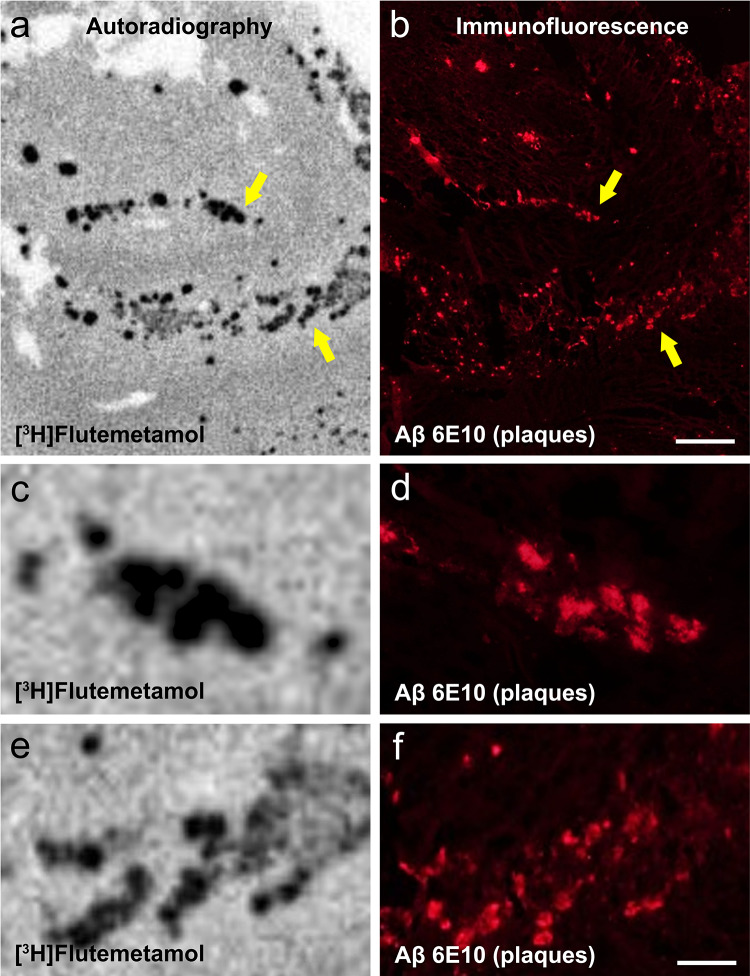
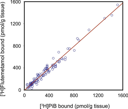
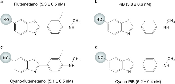
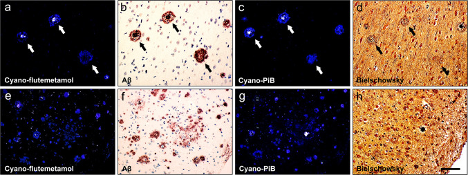
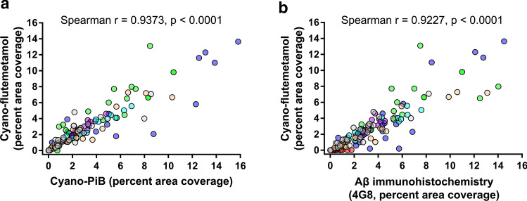
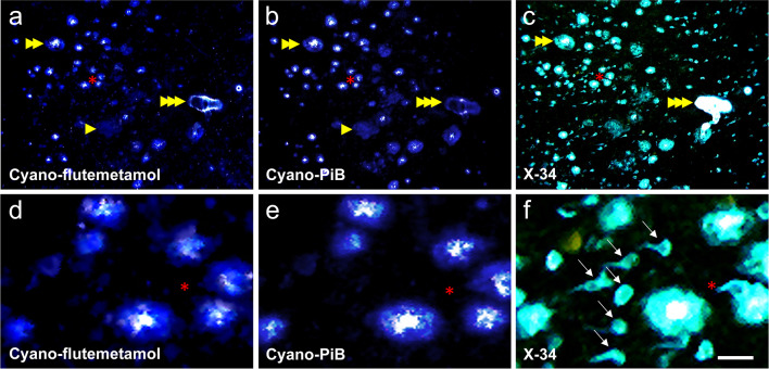
Specificity and sensitivity of positron emission tomography (PET) radiopharmaceuticals targeting fibrillar amyloid-β (Aβ) deposits is high for detection of neuritic Aβ plaques, a mature form of Aβ deposits which often have dense Aβ core (i.e., cored plaques). However, imaging-to-autopsy validation studies of amyloid PET radioligands have identified several false positive cases all of which had mainly diffuse Aβ plaques (i.e., plaques without neuritic pathology or dense amyloid core), and high amyloid PET signal was reported in the striatum where diffuse plaques predominate in Alzheimer's disease (AD). Relative contributions of different plaque types to amyloid PET signal is unclear, particularly in neocortical areas where they are intermixed in AD. In vitro binding assay and autoradiography were performed using [H]flutemetamol and [H]Pittsburgh Compound-B (PiB) in frozen brain homogenates from 30 autopsy cases including sporadic AD and non-AD controls with a range of brain Aβ burden and plaque density. Fixed tissue sections of frontal cortex and caudate from 10 of the AD cases were processed for microscopy using fluorescent derivatives of flutemetamol (cyano-flutemetamol) and PiB (cyano-PiB) and compared to Aβ immunohistochemistry and pan-amyloid (X-34) histology. Using epifluorescence microscopy, percent area coverage and fluorescence output values of cyano-PiB- and cyano-flutemetamol-labeled plaques in two-dimensional microscopic fields were then calculated and combined to obtain integrated density measurements. Using confocal microscopy, we analysed total fluorescence output of the entire three-dimensional volume of individual cored plaques and diffuse plaques labeled with cyano-flutemetamol or cyano-PiB. [H]Flutemetamol and [H]PiB binding values in tissue homogenates correlated strongly and their binding pattern in tissue sections, as seen on autoradiograms, overlapped the pattern of Aβ-immunoreactive plaques on directly adjacent sections. Cyano-flutemetamol and cyano-PiB fluorescence was prominent in cored plaques and less so in diffuse plaques. Across brain regions and cases, percent area coverage of cyano-flutemetamol-labeled plaques correlated strongly with cyano-PiB-labeled and Aβ-immunoreactive plaques. For both ligands, plaque burden, calculated as percent area coverage of all Aβ plaque types, was similar in frontal cortex and caudate regions, while integrated density values were significantly greater in frontal cortex, which contained both cored plaques and diffuse plaques, compared to the caudate, which contained only diffuse plaques. Three-dimensional analysis of individual plaques labeled with either ligand showed that total fluorescence output of a single cored plaque was equivalent to total fluorescence output of approximately three diffuse plaques of similar volume. Our results indicate that [F]flutemetamol and [C]PiB PET signal is influenced by both diffuse plaques and cored plaques, and therefore is likely a function of plaque size and density of Aβ fibrils in plaques. Brain areas with large volumes/frequencies of diffuse plaques could yield [F]flutemetamol and [C]PiB PET retention levels comparable to brain regions with a lower volume/frequency of cored plaques.
正电子发射断层扫描(PET)放射性药物针对纤维状淀粉样β(Aβ)沉积物的特异性和敏感性很高,可用于检测神经原纤维 Aβ斑块,这是 Aβ沉积物的成熟形式,通常具有密集的 Aβ核心(即有核斑块)。然而,淀粉样 PET 配体的成像-尸检验证研究已经确定了几个假阳性病例,这些病例都主要具有弥漫性 Aβ斑块(即没有神经原纤维病理学或密集 Aβ核心的斑块),并且在纹状体中报告了高淀粉样 PET 信号,在那里弥漫性斑块在阿尔茨海默病(AD)中占主导地位。不同斑块类型对淀粉样 PET 信号的相对贡献尚不清楚,特别是在 AD 中斑块相互混合的新皮质区域。使用 [H]flutemetamol 和 [H]Pittsburgh 化合物-B(PiB)在包括散发性 AD 和非 AD 对照的 30 例尸检病例的冷冻脑匀浆中进行了体外结合测定和放射自显影,这些病例的脑 Aβ负担和斑块密度范围广泛。从 10 例 AD 病例的额皮质和尾状核固定组织切片中,使用 flutemetamol(氰基-flutemetamol)和 PiB(氰基-PiB)的荧光衍生物进行显微镜处理,并与 Aβ免疫组织化学和全淀粉样(X-34)组织学进行比较。使用荧光显微镜,然后计算二维显微镜视野中氰基-PiB 和氰基-flutemetamol 标记斑块的面积覆盖率和荧光输出值,并将其组合以获得积分密度测量值。使用共聚焦显微镜,我们分析了用氰基-flutemetamol 或氰基-PiB 标记的单个有核斑块和弥漫性斑块的整个三维体积的总荧光输出。组织匀浆中的 [H]flutemetamol 和 [H]PiB 结合值相关性很强,其在组织切片中的结合模式(如放射自显影所见)与直接相邻切片中 Aβ-免疫反应性斑块的模式重叠。氰基-flutemetamol 和氰基-PiB 荧光在有核斑块中明显,在弥漫性斑块中则不明显。在整个大脑区域和病例中,氰基-flutemetamol 标记斑块的面积覆盖率与氰基-PiB 标记和 Aβ-免疫反应性斑块高度相关。对于两种配体,所有 Aβ 斑块类型的面积覆盖率计算的斑块负担在额皮质和尾状核区域相似,而整合密度值在包含有核斑块和弥漫性斑块的额皮质显著更高,与仅包含弥漫性斑块的尾状核相比。用任一配体标记的单个斑块的三维分析表明,单个有核斑块的总荧光输出相当于类似体积的三个弥漫性斑块的总荧光输出。我们的结果表明,[F]flutemetamol 和 [C]PiB PET 信号受弥漫性斑块和有核斑块的影响,因此可能是 Aβ纤维斑块的大小和密度的函数。具有大体积/弥漫性斑块频率的脑区可能会产生与具有较低体积/有核斑块频率的脑区相当的 [F]flutemetamol 和 [C]PiB PET 保留水平。