Hedlund Eva, Pruszak Jan, Lardaro Thomas, Ludwig Wesley, Viñuela Angel, Kim Kwang-Soo, Isacson Ole
Udall Parkinson's Disease Research Center for Excellence, McLean Hospital, Harvard Medical School, Belmont, Massachusetts 02478, USA.
Stem Cells. 2008 Jun;26(6):1526-36. doi: 10.1634/stemcells.2007-0996. Epub 2008 Apr 3.
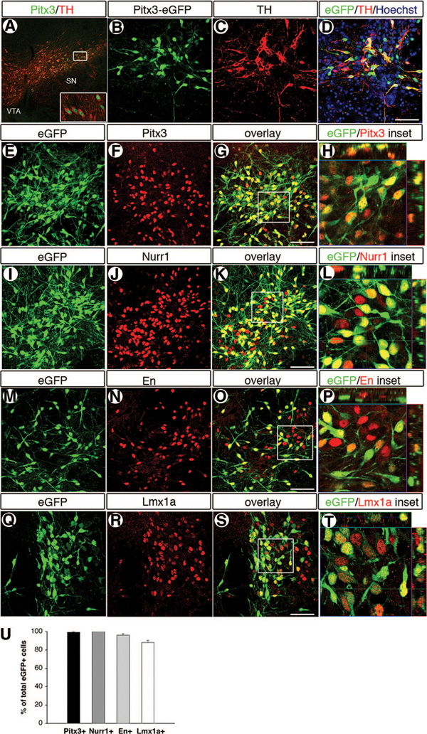
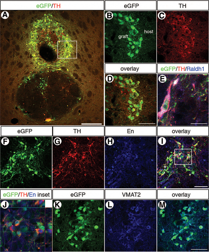
Both fetal ventral mesencephalic (VM) and embryonic stem (ES) cell-derived dopamine neurons have been used successfully to correct behavioral responses in animal models of Parkinson's disease. However, grafts derived from fetal VM cells or from ES cells contain multiple cell types, and the majority of these cells are not dopamine neurons. Isolation of ES cell-derived dopamine neurons and subsequent transplantation would both elucidate the capacity of these neurons to provide functional input and also further explore an efficient and safer use of ES cells for the treatment of Parkinson's disease. Toward this goal, we used a Pitx3-enhanced green fluorescent protein (Pitx3-eGFP) knock-in mouse blastocyst-derived embryonic stem (mES) cell line and fluorescence-activated cell sorting (FACS) to select and purify midbrain dopamine neurons. Initially, the dopaminergic marker profile of intact Pitx3-eGFP mES cultures was evaluated after differentiation in vitro. eGFP expression overlapped closely with that of Pitx3, Nurr1, Engrailed-1, Lmx1a, tyrosine hydroxylase (TH), l-aromatic amino acid decarboxylase (AADC), and vesicular monoamine transporter 2 (VMAT2), demonstrating that these cells were of a midbrain dopamine neuron character. Furthermore, postmitotic Pitx3-eGFP(+) dopamine neurons, which constituted 2%-5% of all live cells in the culture after dissociation, could be highly enriched to >90% purity by FACS, and these isolated neurons were viable, extended neurites, and maintained a dopaminergic profile in vitro. Transplantation to 6-hydroxydopamine-lesioned rats showed that an enriched dopaminergic population could survive and restore both amphetamine- and apomorphine-induced functions, and the grafts contained large numbers of midbrain dopamine neurons, which innervated the host striatum. Disclosure of potential conflicts of interest is found at the end of this article.
胎儿腹侧中脑(VM)和胚胎干细胞(ES)来源的多巴胺能神经元已成功用于纠正帕金森病动物模型中的行为反应。然而,胎儿VM细胞或ES细胞来源的移植物包含多种细胞类型,其中大多数细胞不是多巴胺能神经元。分离ES细胞来源的多巴胺能神经元并随后进行移植,既能阐明这些神经元提供功能性输入的能力,也能进一步探索更有效、更安全地利用ES细胞治疗帕金森病的方法。为实现这一目标,我们使用了一种Pitx3增强绿色荧光蛋白(Pitx3-eGFP)基因敲入小鼠囊胚来源的胚胎干细胞(mES)系和荧光激活细胞分选(FACS)技术来选择和纯化中脑多巴胺能神经元。最初,在体外分化后评估完整的Pitx3-eGFP mES培养物的多巴胺能标记谱。eGFP表达与Pitx3、Nurr1、Engrailed-1、Lmx1a、酪氨酸羟化酶(TH)、L-芳香族氨基酸脱羧酶(AADC)和囊泡单胺转运体2(VMAT2)的表达密切重叠,表明这些细胞具有中脑多巴胺能神经元特征。此外,有丝分裂后的Pitx3-eGFP(+)多巴胺能神经元,在解离后的培养物中占所有活细胞的2%-5%,通过FACS可高度富集至纯度>90%,这些分离的神经元具有活力,能长出神经突,并在体外维持多巴胺能特征。移植到6-羟基多巴胺损伤的大鼠体内表明,富集的多巴胺能群体能够存活并恢复苯丙胺和阿扑吗啡诱导的功能,移植物中含有大量支配宿主纹状体的中脑多巴胺能神经元。潜在利益冲突的披露见本文末尾。