文献检索文档翻译深度研究
Suppr Zotero 插件Zotero 插件
邀请有礼套餐&价格历史记录

新学期,新优惠

限时优惠:9月1日-9月22日

30天高级会员仅需29元

1天体验卡首发特惠仅需5.99元

了解详情
不再提醒
插件&应用
Suppr Zotero 插件Zotero 插件浏览器插件Mac 客户端Windows 客户端微信小程序
高级版
套餐订阅购买积分包
AI 工具
文献检索文档翻译深度研究
关于我们
关于 Suppr公司介绍联系我们用户协议隐私条款
关注我们

Suppr 超能文献

核心技术专利:CN118964589B侵权必究
粤ICP备2023148730 号-1Suppr @ 2025

原发性和转移性黑色素瘤的基因表达谱产生了肿瘤进展和转移的一个转变点。

The gene expression profiles of primary and metastatic melanoma yields a transition point of tumor progression and metastasis.

作者信息

Riker Adam I, Enkemann Steven A, Fodstad Oystein, Liu Suhu, Ren Suping, Morris Christopher, Xi Yaguang, Howell Paul, Metge Brandon, Samant Rajeev S, Shevde Lalita A, Li Wenbin, Eschrich Steven, Daud Adil, Ju Jingfang, Matta Jaime

机构信息

Mitchell Cancer Institute-University of South Alabama, 315 North University Boulevard, MSB 2015, Mobile, Alabama 36688, USA.

出版信息

BMC Med Genomics. 2008 Apr 28;1:13. doi: 10.1186/1755-8794-1-13.


DOI:10.1186/1755-8794-1-13
PMID:18442402
原文链接:https://pmc.ncbi.nlm.nih.gov/articles/PMC2408576/
Abstract

BACKGROUND: The process of malignant transformation, progression and metastasis of melanoma is poorly understood. Gene expression profiling of human cancer has allowed for a unique insight into the genes that are involved in these processes. Thus, we have attempted to utilize this approach through the analysis of a series of primary, non-metastatic cutaneous tumors and metastatic melanoma samples. METHODS: We have utilized gene microarray analysis and a variety of molecular techniques to compare 40 metastatic melanoma (MM) samples, composed of 22 bulky, macroscopic (replaced) lymph node metastases, 16 subcutaneous and 2 distant metastases (adrenal and brain), to 42 primary cutaneous cancers, comprised of 16 melanoma, 11 squamous cell, 15 basal cell skin cancers. A Human Genome U133 Plus 2.0 array from Affymetrix, Inc. was utilized for each sample. A variety of statistical software, including the Affymetrix MAS 5.0 analysis software, was utilized to compare primary cancers to metastatic melanomas. Separate analyses were performed to directly compare only primary melanoma to metastatic melanoma samples. The expression levels of putative oncogenes and tumor suppressor genes were analyzed by semi- and real-time quantitative RT-PCR (qPCR) and Western blot analysis was performed on select genes. RESULTS: 
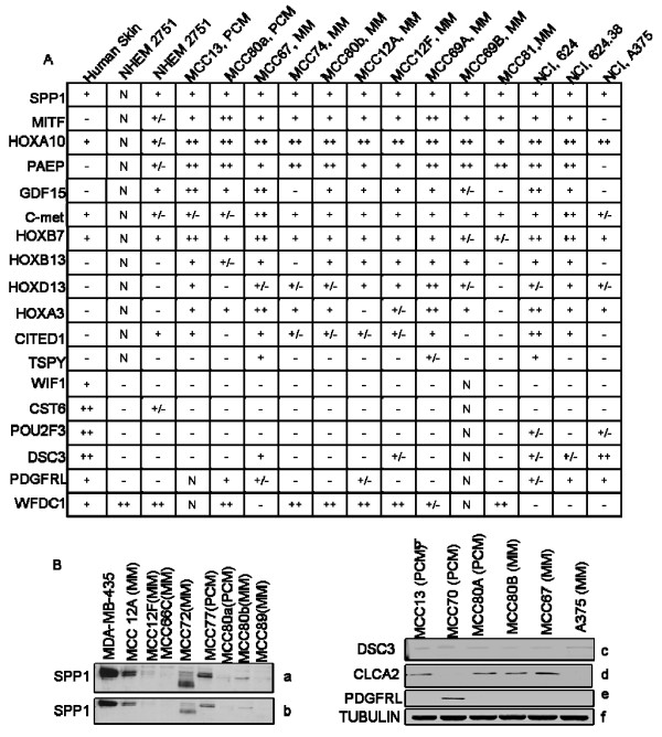
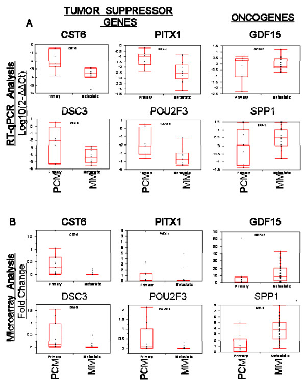
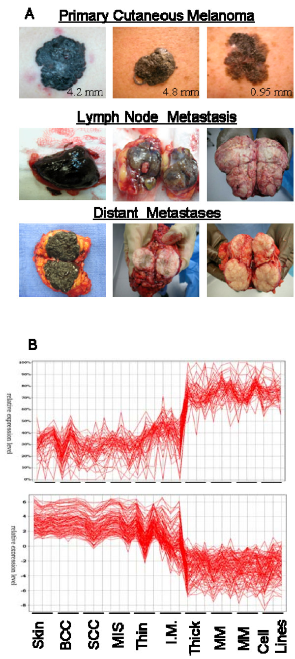
We find that primary basal cell carcinomas, squamous cell carcinomas and thin melanomas express dramatically higher levels of many genes, including SPRR1A/B, KRT16/17, CD24, LOR, GATA3, MUC15, and TMPRSS4, than metastatic melanoma. In contrast, the metastatic melanomas express higher levels of genes such as MAGE, GPR19, BCL2A1, MMP14, SOX5, BUB1, RGS20, and more. The transition from non-metastatic expression levels to metastatic expression levels occurs as melanoma tumors thicken. We further evaluated primary melanomas of varying Breslow's tumor thickness to determine that the transition in expression occurs at different thicknesses for different genes suggesting that the "transition zone" represents a critical time for the emergence of the metastatic phenotype. Several putative tumor oncogenes (SPP-1, MITF, CITED-1, GDF-15, c-Met, HOX loci) and suppressor genes (PITX-1, CST-6, PDGFRL, DSC-3, POU2F3, CLCA2, ST7L), were identified and validated by quantitative PCR as changing expression during this transition period. These are strong candidates for genes involved in the progression or suppression of the metastatic phenotype. CONCLUSION: The gene expression profiling of primary, non-metastatic cutaneous tumors and metastatic melanoma has resulted in the identification of several genes that may be centrally involved in the progression and metastatic potential of melanoma. This has very important implications as we continue to develop an improved understanding of the metastatic process, allowing us to identify specific genes for prognostic markers and possibly for targeted therapeutic approaches.

摘要

背景:黑色素瘤的恶性转化、进展和转移过程目前仍知之甚少。人类癌症的基因表达谱分析使我们能够深入了解参与这些过程的基因。因此,我们试图通过分析一系列原发性、非转移性皮肤肿瘤和转移性黑色素瘤样本,来运用这种方法。 方法:我们利用基因微阵列分析和多种分子技术,将40个转移性黑色素瘤(MM)样本与42个原发性皮肤癌样本进行比较。转移性黑色素瘤样本包括22个体积较大、肉眼可见(替代型)的淋巴结转移灶、16个皮下转移灶和2个远处转移灶(肾上腺和脑转移);原发性皮肤癌样本包括16个黑色素瘤、11个鳞状细胞癌和15个基底细胞皮肤癌。每个样本均使用Affymetrix公司的人类基因组U133 Plus 2.0芯片。使用包括Affymetrix MAS 5.0分析软件在内的多种统计软件,将原发性癌症与转移性黑色素瘤进行比较。单独进行分析以直接比较原发性黑色素瘤与转移性黑色素瘤样本。通过半定量和实时定量逆转录聚合酶链反应(qPCR)分析假定的癌基因和肿瘤抑制基因的表达水平,并对选定基因进行蛋白质免疫印迹分析。 结果:我们发现,原发性基底细胞癌、鳞状细胞癌和薄型黑色素瘤表达许多基因的水平显著高于转移性黑色素瘤,这些基因包括SPRR1A/B、KRT16/17、CD24、LOR、GATA3、MUC15和TMPRSS4等。相比之下,转移性黑色素瘤表达较高水平的基因如MAGE、GPR19、BCL2A1、MMP14、SOX5、BUB1、RGS20等。随着黑色素瘤肿瘤增厚,其表达水平从非转移性转变为转移性。我们进一步评估了不同Breslow肿瘤厚度的原发性黑色素瘤,以确定不同基因在不同厚度时发生表达转变,这表明“转变区”是转移表型出现的关键时期。通过定量PCR鉴定并验证了几个假定的肿瘤癌基因(SPP-1、MITF、CITED-1、GDF-15、c-Met、HOX基因座)和抑制基因(PITX-1、CST-6、PDGFRL、DSC-3、POU2F-组蛋白脱乙酰基酶、CLCA2、ST7L)在这一转变期表达发生变化。这些是参与转移表型进展或抑制的基因的有力候选者。 结论:原发性、非转移性皮肤肿瘤和转移性黑色素瘤的基因表达谱分析已鉴定出几个可能在黑色素瘤进展和转移潜能中起核心作用的基因。这具有非常重要的意义,因为我们继续深入了解转移过程,使我们能够确定用于预后标志物的特定基因,并可能用于靶向治疗方法。

https://cdn.ncbi.nlm.nih.gov/pmc/blobs/6e87/2408576/373bd1c63903/1755-8794-1-13-3.jpg
https://cdn.ncbi.nlm.nih.gov/pmc/blobs/6e87/2408576/39a77017ce23/1755-8794-1-13-1.jpg
https://cdn.ncbi.nlm.nih.gov/pmc/blobs/6e87/2408576/fe5358274f79/1755-8794-1-13-2.jpg
https://cdn.ncbi.nlm.nih.gov/pmc/blobs/6e87/2408576/373bd1c63903/1755-8794-1-13-3.jpg
https://cdn.ncbi.nlm.nih.gov/pmc/blobs/6e87/2408576/39a77017ce23/1755-8794-1-13-1.jpg
https://cdn.ncbi.nlm.nih.gov/pmc/blobs/6e87/2408576/fe5358274f79/1755-8794-1-13-2.jpg
https://cdn.ncbi.nlm.nih.gov/pmc/blobs/6e87/2408576/373bd1c63903/1755-8794-1-13-3.jpg

相似文献

[1]
The gene expression profiles of primary and metastatic melanoma yields a transition point of tumor progression and metastasis.

BMC Med Genomics. 2008-4-28

[2]
Differential gene expression profiling of primary cutaneous melanoma and sentinel lymph node metastases.

Mod Pathol. 2012-3-9

[3]
Mining gene expression signature for the detection of pre-malignant melanocytes and early melanomas with risk for metastasis.

PLoS One. 2012-9-11

[4]
Integrative genome comparison of primary and metastatic melanomas.

PLoS One. 2010-5-24

[5]
Deregulation of glyceraldehyde-3-phosphate dehydrogenase expression during tumor progression of human cutaneous melanoma.

Anticancer Res. 2015-1

[6]
Gene expression profiling of primary cutaneous melanoma and clinical outcome.

J Natl Cancer Inst. 2006-4-5

[7]
Expression of MAGE genes in ocular melanoma during progression from primary to metastatic disease.

Clin Exp Metastasis. 1997-9

[8]
Increased HOX C13 expression in metastatic melanoma progression.

J Transl Med. 2012-5-14

[9]
Early contribution of germline and nevi genetic alterations to a rapidly-progressing cutaneous melanoma patient: a case report.

BMC Med Genomics. 2023-1-5

[10]
Network-based co-expression analysis for exploring the potential diagnostic biomarkers of metastatic melanoma.

PLoS One. 2018-1-29

引用本文的文献

[1]
Multi-Omics Integration with Machine Learning and Molecular Docking Reveals Crosstalk Mechanisms and Drug Candidates in Metastatic Melanoma and Vitiligo.

Clin Cosmet Investig Dermatol. 2025-8-29

[2]
A 3D genome compendium of breast cancer progression.

iScience. 2025-8-5

[3]
Integrated Meta-Analysis Identifies Keratin Family Genes and Associated Genes as Key Biomarkers and Therapeutic Targets in Metastatic Cutaneous Melanoma.

Diagnostics (Basel). 2025-7-13

[4]
Melanoma microsatellites exhibit a metastatic signature by spatial transcriptomics and overexpress mediators of immune evasion.

Virchows Arch. 2025-7-28

[5]
Desmocollin-3 and Bladder Cancer.

Diseases. 2025-4-23

[6]
Analysis of immune infiltration in basal cell carcinoma based on transcriptome sequencing and experimental validation of diagnostic biomarkers.

Discov Oncol. 2025-5-23

[7]
ZNF224 enhances the oncogenic function of p21 via p53 and AKT pathways in melanoma.

FEBS J. 2025-8

[8]
MUC15 acts as a tumor suppressor gene which correlates with prognosis and immune infiltration in esophageal squamous cell carcinoma.

Int J Med Sci. 2025-3-21

[9]
A Novel Butyrate Derivative, Zinc Dibutyroyllysinate, Blunts Microphthalmia-Associated Transcription Factor Expression and Up-Regulates Retinol and Differentiation Pathway mRNAs in a Full-Thickness Human Skin Model.

Int J Mol Sci. 2025-3-9

[10]
The Use of Biologics for Targeting GPCRs in Metastatic Cancers.

BioTech (Basel). 2025-1-30

本文引用的文献

[1]
Systematic search for the best gene expression markers for melanoma micrometastasis detection.

J Pathol. 2007-10

[2]
A high-throughput study in melanoma identifies epithelial-mesenchymal transition as a major determinant of metastasis.

Cancer Res. 2007-4-1

[3]
Gene expression signatures for tumor progression, tumor subtype, and tumor thickness in laser-microdissected melanoma tissues.

Clin Cancer Res. 2007-2-1

[4]
Cancer statistics, 2007.

CA Cancer J Clin. 2007

[5]
Down-regulation of pro-apoptotic genes is an early event in the progression of malignant melanoma.

Ann Surg Oncol. 2007-4

[6]
Decreased PITX1 homeobox gene expression in human lung cancer.

Lung Cancer. 2007-3

[7]
Epigenetic silencing of novel tumor suppressors in malignant melanoma.

Cancer Res. 2006-12-1

[8]
A gene expression signature associated with survival in metastatic melanoma.

J Transl Med. 2006-11-27

[9]
DNA methylation-dependent silencing of CST6 in human breast cancer cell lines.

Lab Invest. 2006-12

[10]
Epigenetic silencing of the tumor suppressor cystatin M occurs during breast cancer progression.

Cancer Res. 2006-8-15

文献AI研究员

20分钟写一篇综述,助力文献阅读效率提升50倍

立即体验

用中文搜PubMed

大模型驱动的PubMed中文搜索引擎

马上搜索

推荐工具

医学文档翻译智能文献检索