Kim Yongbok, Hsu I-Chow J, Pouliot Jean
Department of Radiation Oncology, University of California-San Francisco, Comprehensive Cancer Center, 1600 Divisadero Street, Suite H1031, San Francisco, CA, 94143-1708.
J Appl Clin Med Phys. 2007 Sep 17;8(4):1-13. doi: 10.1120/jacmp.v8i4.2415.
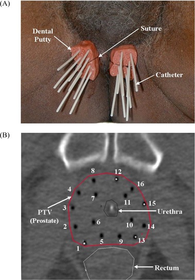
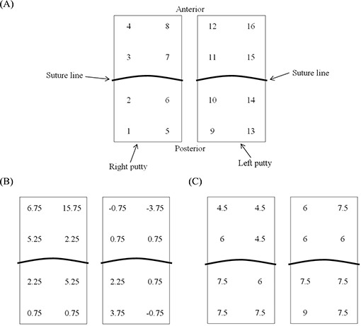
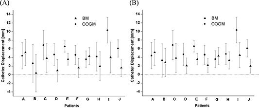
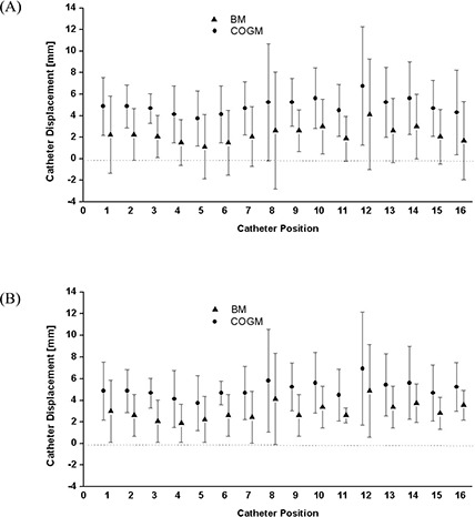
The objective of this work is to measure the cranio-caudal displacement of catheters occurring between consecutive fractions of transrectal ultrasound (TRUS) guided high dose rate (HDR) prostate brachytherapy. Ten consecutive patients were treated with 2 fractions of 9.5 Gy TRUS guided HDR brachytherapy using dental putty for the fixation of catheters. For each patient, a CT scan with 3 mm slice thickness was acquired before each of the two fractions. Two different references were employed to measure the catheter displacement between fractions: the ischial bone as a bony marker (BM) and the center of two gold markers (COGM) implanted in the prostate. The catheter displacement was calculated by multiplying the thickness of CT slice with the difference in number of CT slices between the reference slice and the slice containing the tip of a catheter. The average (range) magnitude of caudal catheter displacement was 2.7 mm (-6.0 to 13.5 mm) for BM method and 5.4 mm (-3.75 to 18.0 mm) for COGM method, respectively. The measurement data obtained from BM and COGM methods verified that both prostate movement and catheter displacement occurred independently between fractions. The most anterior and medial two catheters (catheter position 8 and 12) had the greatest tendency to be displaced in the caudal direction because they were located at the most distant position from the fulcrum, susceptible to the rotation of the dental putty in lateral plane due to the movement of patient legs between fractions. In conclusion, the use of both BM and COGM methods can demonstrate the prostate and catheter movement relative to the BM between fractions. We found a pattern of catheter displacement using our technique. Based on our finding further improvement of our results may be possible by modification of our current technique.
这项工作的目的是测量经直肠超声(TRUS)引导下高剂量率(HDR)前列腺近距离治疗连续分次治疗期间导管的头-尾位移。连续10例患者接受了2次9.5 Gy的TRUS引导下HDR近距离治疗,使用牙科用橡皮泥固定导管。对于每位患者,在两次分次治疗前均进行了层厚为3 mm的CT扫描。采用两种不同的参考点来测量分次治疗间导管的位移:坐骨作为骨标志物(BM)以及植入前列腺的两个金标志物的中心(COGM)。导管位移通过将CT层厚乘以参考层面与包含导管尖端层面之间CT层数的差值来计算。BM法尾端导管位移的平均(范围)幅度分别为2.7 mm(-6.0至13.5 mm),COGM法为5.4 mm(-3.75至18.0 mm)。从BM法和COGM法获得的测量数据证实,前列腺运动和导管位移在分次治疗间是独立发生的。最前侧和中间的两根导管(导管位置8和12)向尾端位移的趋势最大,因为它们位于离支点最远的位置,在分次治疗间由于患者腿部运动,易受牙科用橡皮泥在侧平面旋转的影响。总之,使用BM法和COGM法均可显示分次治疗间前列腺和导管相对于BM的运动。我们利用我们的技术发现了一种导管位移模式。基于我们的发现,通过改进我们目前的技术可能进一步改善我们的结果。