Monk Christopher S, Telzer Eva H, Mogg Karin, Bradley Brendan P, Mai Xiaoqin, Louro Hugo M C, Chen Gang, McClure-Tone Erin B, Ernst Monique, Pine Daniel S
Department of Psychology, University of Michigan, 530 Church St, Ann Arbor, MI 48109-1043, USA.
Arch Gen Psychiatry. 2008 May;65(5):568-76. doi: 10.1001/archpsyc.65.5.568.
Vigilance for threat is a key feature of generalized anxiety disorder (GAD). The amygdala and the ventrolateral prefrontal cortex constitute a neural circuit that is responsible for detection of threats. Disturbed interactions between these structures may underlie pediatric anxiety. To date, no study has selectively examined responses to briefly presented threats in GAD or in pediatric anxiety.
To investigate amygdala and ventrolateral prefrontal cortex activation during processing of briefly presented threats in pediatric GAD.
Case-control study.
Government clinical research institute.
Youth volunteers, 17 with GAD and 12 without a psychiatric diagnosis.
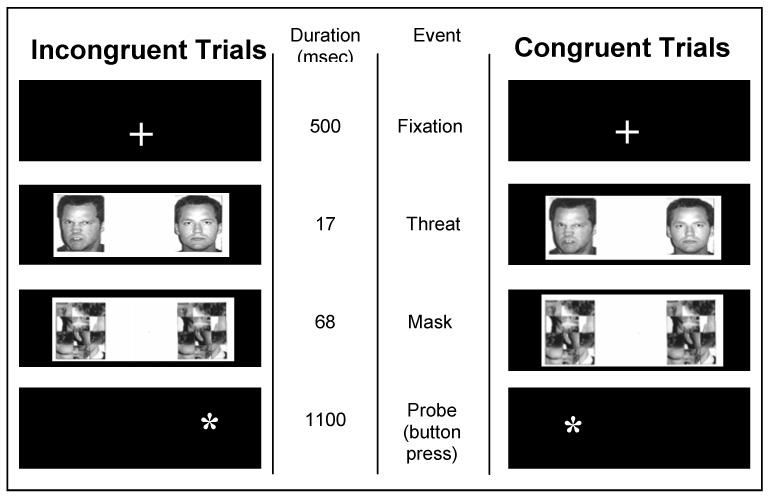
We used functional magnetic resonance imaging to measure blood oxygenation level-dependent signal. During imaging, subjects performed an attention-orienting task with rapidly presented (17 milliseconds) masked emotional (angry or happy) and neutral faces.
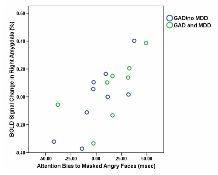
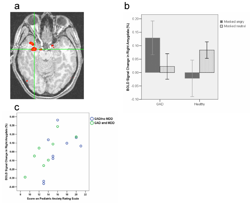
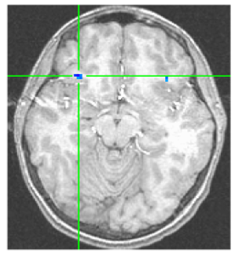
When viewing masked angry faces, youth with GAD relative to comparison subjects showed greater right amygdala activation that positively correlated with anxiety disorder severity. Moreover, in a functional connectivity (psychophysiological interaction) analysis, the right amygdala and the right ventrolateral prefrontal cortex showed strong negative coupling specifically to masked angry faces. This negative coupling tended to be weaker in youth with GAD than in comparison subjects.
Youth with GAD have hyperactivation of the amygdala to briefly presented masked threats. The presence of threat-related negative connectivity between the right ventrolateral prefrontal cortex and the amygdala suggests that the prefrontal cortex modulates the amygdala response to threat. In pediatric GAD, amygdala hyperresponse occurs in the absence of a compensatory increase in modulation by the ventrolateral prefrontal cortex.
对威胁保持警惕是广泛性焦虑症(GAD)的一个关键特征。杏仁核和腹外侧前额叶皮层构成了一个负责检测威胁的神经回路。这些结构之间的相互作用紊乱可能是儿童焦虑症的基础。迄今为止,尚无研究选择性地考察过GAD或儿童焦虑症患者对短暂呈现的威胁的反应。
研究儿童广泛性焦虑症患者在处理短暂呈现的威胁时杏仁核和腹外侧前额叶皮层的激活情况。
病例对照研究。
政府临床研究机构。
青年志愿者,17名患有广泛性焦虑症,12名无精神疾病诊断。
我们使用功能磁共振成像来测量血氧水平依赖信号。在成像过程中,受试者执行一项注意力定向任务,快速呈现(17毫秒)带有掩蔽的情绪化(愤怒或高兴)和中性面孔。
在观看带有掩蔽的愤怒面孔时,与对照组相比,患有广泛性焦虑症的青年右侧杏仁核激活程度更高,且与焦虑症严重程度呈正相关。此外,在功能连接(心理生理交互作用)分析中,右侧杏仁核与右侧腹外侧前额叶皮层对带有掩蔽的愤怒面孔表现出强烈的负耦合。这种负耦合在患有广泛性焦虑症的青年中往往比对照组更弱。
患有广泛性焦虑症的青年在面对短暂呈现的掩蔽威胁时杏仁核过度激活。右侧腹外侧前额叶皮层与杏仁核之间存在与威胁相关的负连接,这表明前额叶皮层调节杏仁核对威胁的反应。在儿童广泛性焦虑症中,杏仁核过度反应发生时,腹外侧前额叶皮层的调节作用并未相应增加。