Strasfeld David B, Ling Yun L, Shim Sang-Hee, Zanni Martin T
Department of Chemistry, University of Wisconsin-Madison, 1101 University Avenue, Madison,Wisconsin 53703, USA.
J Am Chem Soc. 2008 May 28;130(21):6698-9. doi: 10.1021/ja801483n. Epub 2008 May 7.
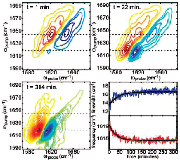
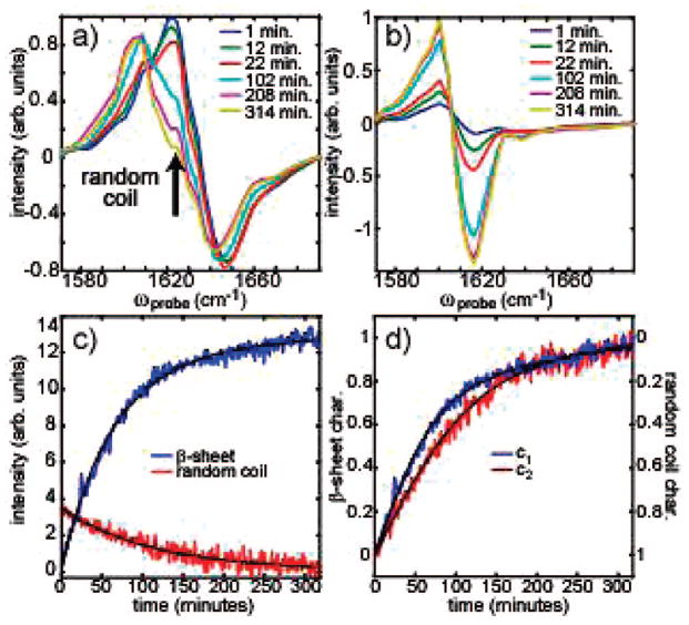
Amyloid forming proteins have been implicated in many human diseases. The kinetics of amyloid fiber formation are of particular interest because evidence points to intermediate folding structures as potential cytotoxic species. The standard methods for monitoring the kinetics are to use fluorescence or circular dichroism spectroscopy, which do not uniquely resolve secondary structures. In this work, we use a new technology for rapidly scanning 2D-IR spectra that allows us to follow the fiber formation kinetics of the human islet amyloid polypeptide (hIAPP) that is involved in type II diabetes. Spectroscopic markers are identified that uniquely monitor random coil versus beta-sheet secondary structures as well as probe beta-sheet elongation and stacking. Our measurements provide more rigorous kinetics for the secondary structure evolution of amyloid formation than is available with other techniques.
淀粉样蛋白形成蛋白与许多人类疾病有关。淀粉样纤维形成的动力学尤其令人感兴趣,因为有证据表明中间折叠结构是潜在的细胞毒性物质。监测动力学的标准方法是使用荧光或圆二色光谱法,但这些方法无法唯一地解析二级结构。在这项工作中,我们使用了一种用于快速扫描二维红外光谱的新技术,该技术使我们能够追踪参与II型糖尿病的人胰岛淀粉样多肽(hIAPP)的纤维形成动力学。我们鉴定出了光谱标记物,它们能够唯一地监测无规卷曲与β-折叠二级结构,同时还能探测β-折叠的延伸和堆积。与其他技术相比,我们的测量为淀粉样蛋白形成过程中二级结构的演变提供了更精确的动力学数据。