• 文献检索
  • 文档翻译
  • 深度研究
  • 学术资讯
  • Suppr Zotero 插件Zotero 插件
  • 邀请有礼
  • 套餐&价格
  • 历史记录
应用&插件
Suppr Zotero 插件Zotero 插件浏览器插件Mac 客户端Windows 客户端微信小程序
定价
高级版会员购买积分包购买API积分包
服务
文献检索文档翻译深度研究API 文档MCP 服务
关于我们
关于 Suppr公司介绍联系我们用户协议隐私条款
关注我们

Suppr 超能文献

核心技术专利:CN118964589B侵权必究
粤ICP备2023148730 号-1Suppr @ 2026

文献检索

告别复杂PubMed语法,用中文像聊天一样搜索,搜遍4000万医学文献。AI智能推荐,让科研检索更轻松。

立即免费搜索

文件翻译

保留排版,准确专业,支持PDF/Word/PPT等文件格式,支持 12+语言互译。

免费翻译文档

深度研究

AI帮你快速写综述,25分钟生成高质量综述,智能提取关键信息,辅助科研写作。

立即免费体验

小鼠逆转录病毒诱导的海绵状脊髓脑病:感染靶细胞的区域分布以及在神经元无病毒表达情况下发生的神经元丢失。

Retrovirus-induced spongiform myeloencephalopathy in mice: regional distribution of infected target cells and neuronal loss occurring in the absence of viral expression in neurons.

作者信息

Kay D G, Gravel C, Robitaille Y, Jolicoeur P

机构信息

Laboratory of Molecular Biology, Clinical Research Institute of Montreal, Canada.

出版信息

Proc Natl Acad Sci U S A. 1991 Feb 15;88(4):1281-5. doi: 10.1073/pnas.88.4.1281.

DOI:10.1073/pnas.88.4.1281
PMID:1847516
原文链接:https://pmc.ncbi.nlm.nih.gov/articles/PMC51001/
Abstract

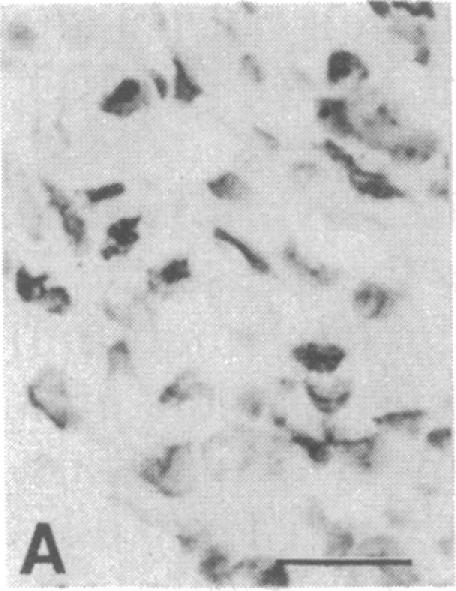
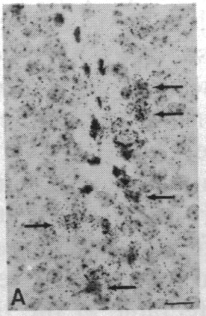
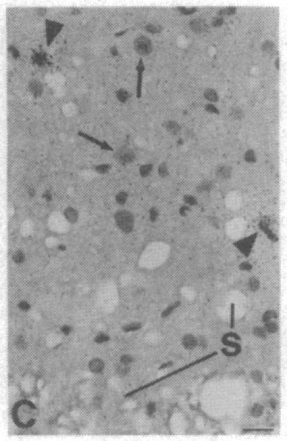
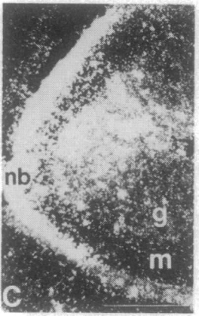
The Cas-Br-E murine leukemia virus (MuLV) induces a spongiform myeloencephalopathy resulting in a progressive hindlimb paralysis. We have used in situ hybridization with a Cas-Br-E MuLV-specific probe to study viral expression in the central nervous system. Infected cells were concentrated in regions where spongiform lesions and gliosis are detected (lumbosacral spinal cord, brainstem, deep cerebellar regions), suggesting a causative link between the level of virus expression and the degree of pathological changes in this disease. However, viral expression was not in itself sufficient to cause disease, since significant viral expression was observed in regions that did not exhibit pathological changes (cerebellar cortex, hippocampus, corpus callosum, peripheral nervous system). In both diseased and nondiseased regions, endothelial and glial cells were identified as the main target cells. Neurons in diseased regions did not show viral expression. The regional distribution of the spongiform changes appears to be laid down very early following infection, since expression could be detected at 10 days postinfection in regions that become diseased. These results indicate that nonneuronal cells have distinct properties in various regions of the central nervous system and suggest an indirect mechanism of neuronal loss consequent to viral expression in nonneuronal cells.

摘要

卡斯 - 布 - 埃氏鼠白血病病毒(MuLV)可引发一种海绵状脊髓脑病,导致后肢进行性麻痹。我们使用了与卡斯 - 布 - 埃氏MuLV特异性探针进行原位杂交,以研究病毒在中枢神经系统中的表达。受感染细胞集中在检测到海绵状病变和胶质增生的区域(腰骶脊髓、脑干、小脑深部区域),这表明病毒表达水平与该疾病的病理变化程度之间存在因果关系。然而,病毒表达本身并不足以引发疾病,因为在未出现病理变化的区域(小脑皮质、海马体、胼胝体、外周神经系统)也观察到了显著的病毒表达。在患病和未患病区域,内皮细胞和神经胶质细胞均被确定为主要靶细胞。患病区域的神经元未显示病毒表达。海绵状变化的区域分布似乎在感染后很早就已确定,因为在感染后10天就可以在患病区域检测到表达。这些结果表明,非神经元细胞在中枢神经系统的各个区域具有不同特性,并提示非神经元细胞中病毒表达导致神经元丢失的间接机制。

https://cdn.ncbi.nlm.nih.gov/pmc/blobs/bd22/51001/3f6c0b6d3a94/pnas01054-0209-j.jpg
https://cdn.ncbi.nlm.nih.gov/pmc/blobs/bd22/51001/d75870f82e2f/pnas01054-0207-a.jpg
https://cdn.ncbi.nlm.nih.gov/pmc/blobs/bd22/51001/ab38a1a42e2a/pnas01054-0207-b.jpg
https://cdn.ncbi.nlm.nih.gov/pmc/blobs/bd22/51001/0bc8aa500224/pnas01054-0207-c.jpg
https://cdn.ncbi.nlm.nih.gov/pmc/blobs/bd22/51001/e28f35a3d8a1/pnas01054-0208-a.jpg
https://cdn.ncbi.nlm.nih.gov/pmc/blobs/bd22/51001/8810c4378d08/pnas01054-0208-b.jpg
https://cdn.ncbi.nlm.nih.gov/pmc/blobs/bd22/51001/df61bf1fa1bd/pnas01054-0208-c.jpg
https://cdn.ncbi.nlm.nih.gov/pmc/blobs/bd22/51001/5ca02f897933/pnas01054-0208-d.jpg
https://cdn.ncbi.nlm.nih.gov/pmc/blobs/bd22/51001/1e8ecbf42e5f/pnas01054-0208-e.jpg
https://cdn.ncbi.nlm.nih.gov/pmc/blobs/bd22/51001/bb2d29033a7d/pnas01054-0208-f.jpg
https://cdn.ncbi.nlm.nih.gov/pmc/blobs/bd22/51001/f941e6807c25/pnas01054-0208-g.jpg
https://cdn.ncbi.nlm.nih.gov/pmc/blobs/bd22/51001/63edb1e664ea/pnas01054-0209-a.jpg
https://cdn.ncbi.nlm.nih.gov/pmc/blobs/bd22/51001/a8e6f5595ac4/pnas01054-0209-b.jpg
https://cdn.ncbi.nlm.nih.gov/pmc/blobs/bd22/51001/624c497982a4/pnas01054-0209-c.jpg
https://cdn.ncbi.nlm.nih.gov/pmc/blobs/bd22/51001/a8a92f525a4a/pnas01054-0209-d.jpg
https://cdn.ncbi.nlm.nih.gov/pmc/blobs/bd22/51001/b21ebd189af9/pnas01054-0209-e.jpg
https://cdn.ncbi.nlm.nih.gov/pmc/blobs/bd22/51001/134d9b67915b/pnas01054-0209-f.jpg
https://cdn.ncbi.nlm.nih.gov/pmc/blobs/bd22/51001/ded442cec8a7/pnas01054-0209-g.jpg
https://cdn.ncbi.nlm.nih.gov/pmc/blobs/bd22/51001/184865052c0c/pnas01054-0209-h.jpg
https://cdn.ncbi.nlm.nih.gov/pmc/blobs/bd22/51001/0e48fbcd8b3b/pnas01054-0209-i.jpg
https://cdn.ncbi.nlm.nih.gov/pmc/blobs/bd22/51001/3f6c0b6d3a94/pnas01054-0209-j.jpg

相似文献

1
Retrovirus-induced spongiform myeloencephalopathy in mice: regional distribution of infected target cells and neuronal loss occurring in the absence of viral expression in neurons.小鼠逆转录病毒诱导的海绵状脊髓脑病:感染靶细胞的区域分布以及在神经元无病毒表达情况下发生的神经元丢失。
Proc Natl Acad Sci U S A. 1991 Feb 15;88(4):1281-5. doi: 10.1073/pnas.88.4.1281.
2
Substitution of the U3 long terminal repeat region of the neurotropic Cas-Br-E retrovirus affects its disease-inducing potential.嗜神经性Cas-Br-E逆转录病毒U3长末端重复序列区域的替换会影响其致病潜力。
J Virol. 1990 Aug;64(8):3742-52. doi: 10.1128/JVI.64.8.3742-3752.1990.
3
Identification of the infected target cell type in spongiform myeloencephalopathy induced by the neurotropic Cas-Br-E murine leukemia virus.嗜神经性卡斯-布-埃氏小鼠白血病病毒诱导的海绵状脑脊髓病中受感染靶细胞类型的鉴定
J Virol. 1993 Nov;67(11):6648-58. doi: 10.1128/JVI.67.11.6648-6658.1993.
4
Development of spongiform encephalopathy in retroviral infected mice.逆转录病毒感染小鼠中海绵状脑病的发展
Lab Invest. 1992 Mar;66(3):292-302.
5
Role of abortive retroviral infection of neurons in spongiform CNS degeneration.神经元的流产性逆转录病毒感染在海绵状中枢神经系统变性中的作用。
Nature. 1990 Jul 12;346(6280):181-3. doi: 10.1038/346181a0.
6
Kinetic analysis of spongiform neurodegenerative disease induced by a highly virulent murine retrovirus.一种高毒力鼠逆转录病毒诱导的海绵状神经退行性疾病的动力学分析
Lab Invest. 1994 May;70(5):711-23.
7
Murine retrovirus-induced spongiform encephalopathy: productive infection of microglia and cerebellar neurons in accelerated CNS disease.小鼠逆转录病毒诱导的海绵状脑病:在加速的中枢神经系统疾病中,小胶质细胞和小脑神经元的生产性感染。
Neuron. 1991 Sep;7(3):365-79. doi: 10.1016/0896-6273(91)90289-c.
8
Temporal central and peripheral nervous system changes induced by a paralytogenic mutant of Moloney murine leukemia virus TB.莫洛尼鼠白血病病毒TB致瘫突变体引起的颞叶中枢和外周神经系统变化
Lab Invest. 1993 Dec;69(6):724-35.
9
Viral load and its relationship to quinolinic acid, TNF alpha, and IL-6 levels in the CNS of retroviral infected mice.
Mol Chem Neuropathol. 1994 Aug;22(3):143-60. doi: 10.1007/BF03160102.
10
Retrovirus-induced murine motor neuron disease: mapping the determinant of spongiform degeneration within the envelope gene.逆转录病毒诱导的小鼠运动神经元疾病:确定包膜基因内海绵状变性的决定因素。
Proc Natl Acad Sci U S A. 1989 May;86(10):3896-900. doi: 10.1073/pnas.86.10.3896.

引用本文的文献

1
Astrocyte Infection Is Required for Retrovirus-Induced Spongiform Neurodegeneration Despite Suppressed Viral Protein Expression.尽管病毒蛋白表达受到抑制,但逆转录病毒诱导的海绵状神经退行性变仍需要星形胶质细胞感染。
Front Neurosci. 2019 Oct 29;13:1166. doi: 10.3389/fnins.2019.01166. eCollection 2019.
2
Ecotropic Murine Leukemia Virus Infection of Glial Progenitors Interferes with Oligodendrocyte Differentiation: Implications for Neurovirulence.嗜亲性鼠白血病病毒对神经胶质前体细胞的感染干扰少突胶质细胞分化:对神经毒力的影响
J Virol. 2016 Jan 13;90(7):3385-99. doi: 10.1128/JVI.03156-15.
3
Postinhibitory rebound neurons and networks are disrupted in retrovirus-induced spongiform neurodegeneration.

本文引用的文献

1
Pathogenesis of the slow disease of the central nervous system associated with wild mouse virus. III. Role of input virus and MCF recombinants in disease.
Virology. 1983 Jul 15;128(1):154-65. doi: 10.1016/0042-6822(83)90326-4.
2
Molecular cloning of infectious viral DNA from ecotropic neurotropic wild mouse retrovirus.从亲嗜性嗜神经性野生小鼠逆转录病毒中克隆感染性病毒DNA
J Virol. 1983 Mar;45(3):1159-63. doi: 10.1128/JVI.45.3.1159-1163.1983.
3
Spongiform polioencephalomyelopathy caused by a murine retrovirus. II. Ultrastructural localization of virus replication and spongiform changes in the central nervous system.由鼠逆转录病毒引起的海绵状脑脊髓灰质炎。II. 病毒复制的超微结构定位及中枢神经系统中的海绵状变化
抑制后反弹神经元和神经网络在逆转录病毒诱导的海绵状神经变性中受到破坏。
J Neurophysiol. 2014 Aug 1;112(3):683-704. doi: 10.1152/jn.00227.2014. Epub 2014 May 14.
4
Unique N-linked glycosylation of CasBrE Env influences its stability, processing, and viral infectivity but not its neurotoxicity.CasBrE 包膜蛋白的独特 N -linked 糖基化影响其稳定性、加工和病毒感染力,但不影响其神经毒性。
J Virol. 2013 Aug;87(15):8372-87. doi: 10.1128/JVI.00392-13. Epub 2013 May 22.
5
Senescence-accelerated Mice (SAMs) as a Model for Brain Aging and Immunosenescence.衰老加速小鼠(SAMs)作为脑衰老和免疫衰老模型。
Aging Dis. 2011 Oct;2(5):414-35. Epub 2011 Oct 28.
6
Poliomyelitis in MuLV-infected ICR-SCID mice after injection of basement membrane matrix contaminated with lactate dehydrogenase-elevating virus.注射了被乳酸脱氢酶升高病毒污染的基底膜基质后,MuLV感染的ICR-SCID小鼠发生的脊髓灰质炎
Comp Med. 2011 Oct;61(5):404-11.
7
Retrovirus-induced spongiform neurodegeneration is mediated by unique central nervous system viral targeting and expression of env alone.逆转录病毒诱导的海绵状神经退行性变是由独特的中枢神经系统病毒靶向和单独表达 env 介导的。
J Virol. 2011 Mar;85(5):2060-78. doi: 10.1128/JVI.02210-10. Epub 2010 Dec 29.
8
The degree of folding instability of the envelope protein of a neurovirulent murine retrovirus correlates with the severity of the neurological disease.一种神经毒性鼠逆转录病毒包膜蛋白的折叠不稳定性程度与神经疾病的严重程度相关。
J Virol. 2009 Jun;83(12):6079-86. doi: 10.1128/JVI.02647-08. Epub 2009 Apr 1.
9
Oligodendrocytes are a major target of the toxicity of spongiogenic murine retroviruses.少突胶质细胞是海绵状鼠逆转录病毒毒性的主要靶点。
Am J Pathol. 2006 Sep;169(3):1026-38. doi: 10.2353/ajpath.2006.051357.
10
Gene expression profiling of microglia infected by a highly neurovirulent murine leukemia virus: implications for neuropathogenesis.高神经毒性小鼠白血病病毒感染的小胶质细胞的基因表达谱分析:对神经发病机制的影响
Retrovirology. 2006 May 12;3:26. doi: 10.1186/1742-4690-3-26.
Neuropathol Appl Neurobiol. 1981 Sep-Oct;7(5):365-80. doi: 10.1111/j.1365-2990.1981.tb00239.x.
4
Pathogenesis of the slow disease of the central nervous system associated with wild mouse virus. II. Role of virus and host gene products.与野生小鼠病毒相关的中枢神经系统慢病毒病的发病机制。II. 病毒和宿主基因产物的作用。
Virology. 1980 Nov;107(1):180-93. doi: 10.1016/0042-6822(80)90283-4.
5
Spongiform polioencephalomyelopathy caused by a murine retrovirus. I. Pathogenesis of infection in newborn mice.由鼠逆转录病毒引起的海绵状脑脊髓灰质炎。I. 新生小鼠感染的发病机制。
Lab Invest. 1980 Nov;43(5):480-6.
6
Physical mapping of the paralysis-inducing determinant of a wild mouse ecotropic neurotropic retrovirus.一种野生小鼠亲嗜性嗜神经性逆转录病毒致瘫决定簇的物理图谱分析
J Virol. 1984 Nov;52(2):356-63. doi: 10.1128/JVI.52.2.356-363.1984.
7
Efficient in vitro synthesis of biologically active RNA and RNA hybridization probes from plasmids containing a bacteriophage SP6 promoter.从含有噬菌体SP6启动子的质粒中高效体外合成生物活性RNA和RNA杂交探针。
Nucleic Acids Res. 1984 Sep 25;12(18):7035-56. doi: 10.1093/nar/12.18.7035.
8
Lower motor neuron degeneration associated with type C RNA virus infection in mice: neuropathological features.小鼠中与C型RNA病毒感染相关的下运动神经元变性:神经病理学特征
J Neuropathol Exp Neurol. 1974 Apr;33(2):285-307. doi: 10.1097/00005072-197404000-00007.
9
A spontaneous lower motor neuron disease apparently caused by indigenous type-C RNA virus in wild mice.一种明显由野生小鼠体内的本土C型RNA病毒引起的自发性下运动神经元疾病。
J Natl Cancer Inst. 1973 Oct;51(4):1243-54. doi: 10.1093/jnci/51.4.1243.
10
Immunohistochemical localization of macrophages and microglia in the adult and developing mouse brain.巨噬细胞和小胶质细胞在成年及发育中小鼠大脑中的免疫组织化学定位
Neuroscience. 1985 Jun;15(2):313-26. doi: 10.1016/0306-4522(85)90215-5.