Chen Huiyi, Liu Bin, Lukas Thomas J, Neufeld Arthur H
Forsythe Laboratory for the Investigation of the Aging Retina, Department of Ophthalmology, Northwestern University School of Medicine, Chicago, Illinois, United States of America.
PLoS One. 2008 Jun 4;3(6):e2339. doi: 10.1371/journal.pone.0002339.
Although the statement that age is the greatest risk factor for Age-related macular degeneration (AMD) is widely accepted, the cellular and molecular explanations for that clinical statement are not generally known. A major focus of AMD research is the retinal pigment epithelium (RPE)/choroid. The purpose of this study was to characterize the changes in the RPE/choroid with age that may provide a background for the development of AMD.
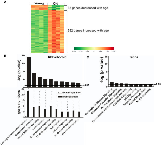
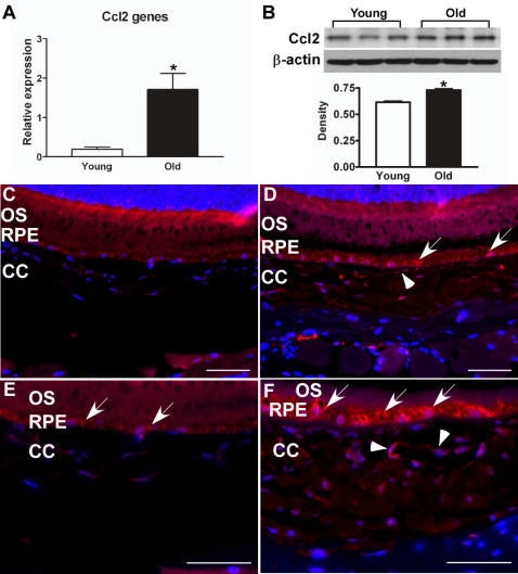
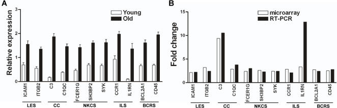
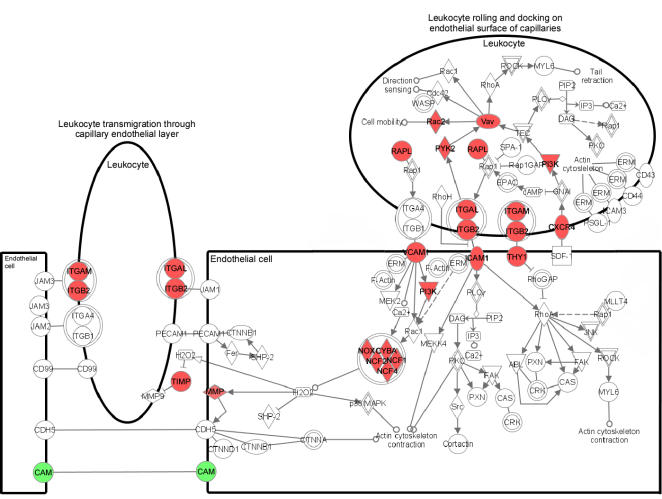
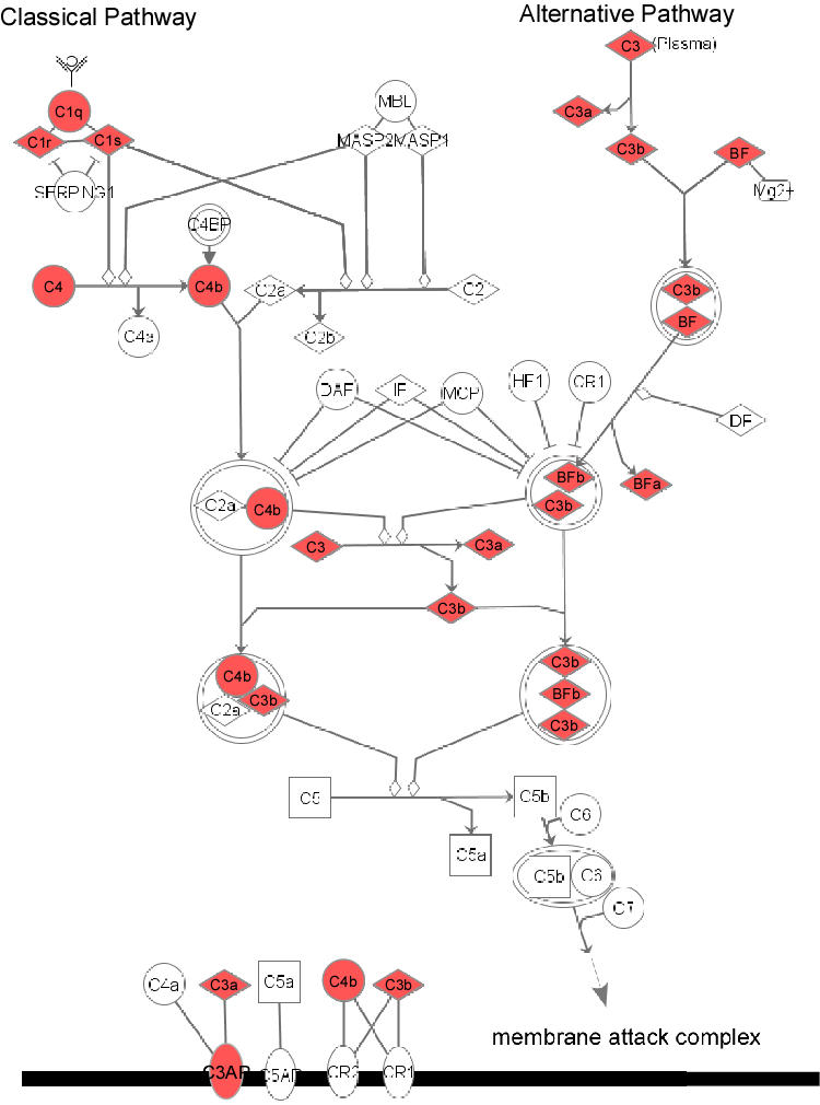
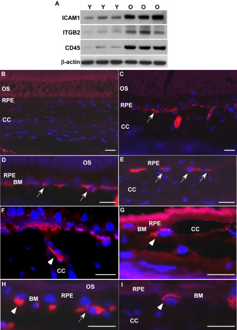
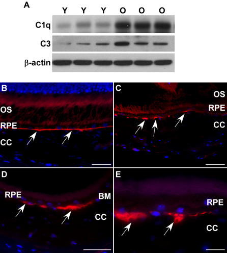
METHODOLOGY/PRINCIPAL FINDINGS: We compared the transcriptional profiles, key protein levels and histology of the RPE/choroid from young and old mice. Using three statistical methods, microarray data demonstrated marked changes in the old mouse. There were 315 genes differentially expressed with age; most of these genes were related to immune responses and inflammatory activity. Canonical pathways having significant numbers of upregulated genes in aged RPE/choroid included leukocyte extravasation, complement cascades, natural killer cell signaling and IL-10 signaling. By contrast, the adjacent neural retina showed completely different age-related changes. The levels of proteins that participate in leukocyte extravasation and complement pathways were consistently increased in the normal, aged RPE/choroid. Furthermore, there was increased gene expression and protein levels of leukocyte attracting signal, chemokine ligand 2 (Ccl2) in aged RPE/choroid. In old animals, there was marked extravasation and accumulation of leukocytes from the choroidal circulation onto Bruch's membrane and into the RPE.
CONCLUSIONS/SIGNIFICANCE: These phenotypic changes indicate that the RPE/choroid in the normal, old mouse has become an immunologically active tissue. There are signals from the normal, aged RPE/choroid which recruit leukocytes from the circulation and activate the complement cascade. These age-related changes that occur in the RPE/choroid with age, to the extent that they occur in the human retina, may provide the background for an error in regulation of immunological activity to cause AMD to appear in an elderly individual.
尽管年龄是年龄相关性黄斑变性(AMD)最大风险因素这一说法已被广泛接受,但该临床说法的细胞和分子学解释却并不为人熟知。AMD研究的一个主要焦点是视网膜色素上皮(RPE)/脉络膜。本研究的目的是描述RPE/脉络膜随年龄增长的变化,这可能为AMD的发生提供背景。
方法/主要发现:我们比较了年轻和老年小鼠RPE/脉络膜的转录谱、关键蛋白水平和组织学。使用三种统计方法,微阵列数据显示老年小鼠有显著变化。有315个基因随年龄差异表达;这些基因大多与免疫反应和炎症活动有关。在老年RPE/脉络膜中上调基因数量较多的典型通路包括白细胞渗出、补体级联反应、自然杀伤细胞信号传导和IL-10信号传导。相比之下,相邻的神经视网膜显示出完全不同的年龄相关变化。参与白细胞渗出和补体途径的蛋白水平在正常老年RPE/脉络膜中持续升高。此外,老年RPE/脉络膜中白细胞吸引信号趋化因子配体2(Ccl2)的基因表达和蛋白水平增加。在老年动物中,有明显的白细胞从脉络膜循环渗出并积聚到Bruch膜上并进入RPE。
结论/意义:这些表型变化表明正常老年小鼠的RPE/脉络膜已成为具有免疫活性的组织。正常老年RPE/脉络膜发出信号,从循环中募集白细胞并激活补体级联反应。RPE/脉络膜随年龄发生的这些与年龄相关的变化,只要它们发生在人类视网膜中,可能为免疫活性调节错误导致老年人出现AMD提供背景。