Casas François, Pessemesse Laurence, Grandemange Stéphanie, Seyer Pascal, Gueguen Naïg, Baris Olivier, Lepourry Laurence, Cabello Gérard, Wrutniak-Cabello Chantal
INRA, UMR866 Différenciation cellulaire et croissance, Montpellier, France.
PLoS One. 2008 Jun 25;3(6):e2501. doi: 10.1371/journal.pone.0002501.
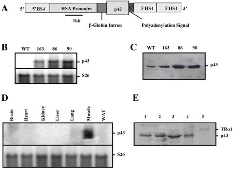
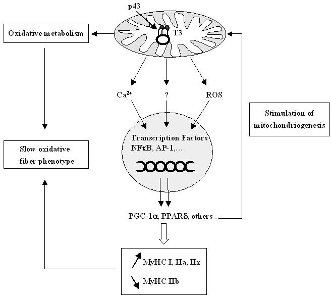
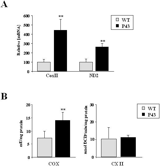
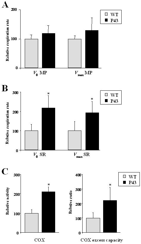
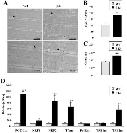
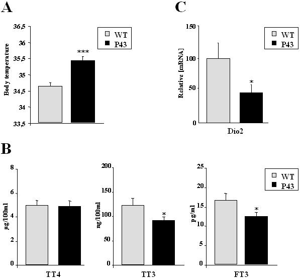
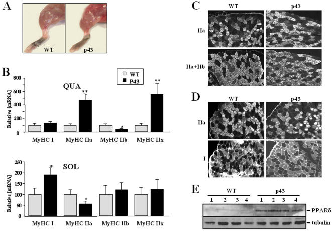
In previous studies, we have characterized a new hormonal pathway involving a mitochondrial T3 receptor (p43) acting as a mitochondrial transcription factor and consequently stimulating mitochondrial activity and mitochondrial biogenesis. We have established the involvement of this T3 pathway in the regulation of in vitro myoblast differentiation. We have generated mice overexpressing p43 under control of the human alpha-skeletal actin promoter. In agreement with the previous characterization of this promoter, northern-blot and western-blot experiments confirmed that after birth p43 was specifically overexpressed in skeletal muscle. As expected from in vitro studies, in 2-month old mice, p43 overexpression increased mitochondrial genes expression and mitochondrial biogenesis as attested by the increase of mitochondrial mass and mt-DNA copy number. In addition, transgenic mice had a body temperature 0.8 degrees C higher than control ones and displayed lower plasma triiodothyronine levels. Skeletal muscles of transgenic mice were redder than wild-type animals suggesting an increased oxidative metabolism. In line with this observation, in gastrocnemius, we recorded a strong increase in cytochrome oxidase activity and in mitochondrial respiration. Moreover, we observed that p43 drives the formation of oxidative fibers: in soleus muscle, where MyHC IIa fibers were partly replaced by type I fibers; in gastrocnemius muscle, we found an increase in MyHC IIa and IIx expression associated with a reduction in the number of glycolytic fibers type IIb. In addition, we found that PGC-1alpha and PPARdelta, two major regulators of muscle phenotype were up regulated in p43 transgenic mice suggesting that these proteins could be downstream targets of mitochondrial activity. These data indicate that the direct mitochondrial T3 pathway is deeply involved in the acquisition of contractile and metabolic features of muscle fibers in particular by regulating PGC-1alpha and PPARdelta.
在先前的研究中,我们已经鉴定出一条新的激素信号通路,该通路涉及一种作为线粒体转录因子的线粒体T3受体(p43),进而刺激线粒体活性和线粒体生物合成。我们已经证实这条T3信号通路参与体外成肌细胞分化的调控。我们构建了在人α-骨骼肌肌动蛋白启动子控制下过表达p43的小鼠。与该启动子先前的特性一致,Northern印迹和Western印迹实验证实,出生后p43在骨骼肌中特异性过表达。正如体外研究预期的那样,在2月龄小鼠中,p43过表达增加了线粒体基因表达和线粒体生物合成,线粒体质量和mt-DNA拷贝数的增加证明了这一点。此外,转基因小鼠的体温比对照小鼠高0.8摄氏度,且血浆三碘甲状腺原氨酸水平较低。转基因小鼠的骨骼肌比野生型动物更红,表明氧化代谢增加。与此观察结果一致,在腓肠肌中,我们记录到细胞色素氧化酶活性和线粒体呼吸显著增加。此外,我们观察到p43驱动氧化纤维的形成:在比目鱼肌中,MyHC IIa纤维部分被I型纤维取代;在腓肠肌中,我们发现MyHC IIa和IIx表达增加,同时IIb型糖酵解纤维数量减少。此外,我们发现肌肉表型的两个主要调节因子PGC-1α和PPARδ在p43转基因小鼠中上调,这表明这些蛋白质可能是线粒体活性的下游靶点。这些数据表明,直接的线粒体T3信号通路尤其通过调节PGC-1α和PPARδ,深度参与肌肉纤维收缩和代谢特征的获得。