Martin Bronwen, Brenneman Randall, Becker Kevin G, Gucek Marjan, Cole Robert N, Maudsley Stuart
Laboratory of Neurosciences, National Institute on Aging Intramural Research Program, Biomedical Research Center, Baltimore, Maryland, United States of America.
PLoS One. 2008 Jul 23;3(7):e2750. doi: 10.1371/journal.pone.0002750.
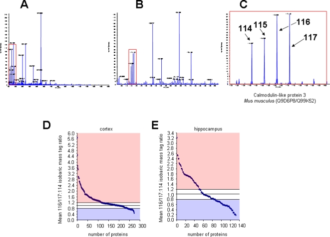
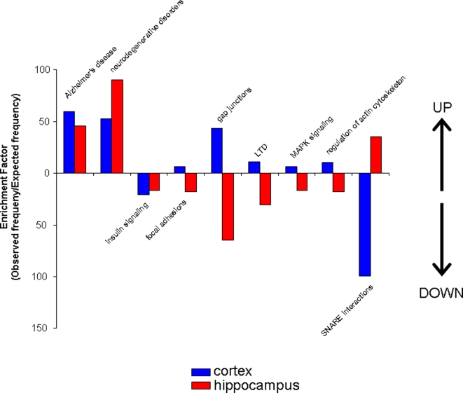
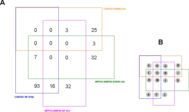
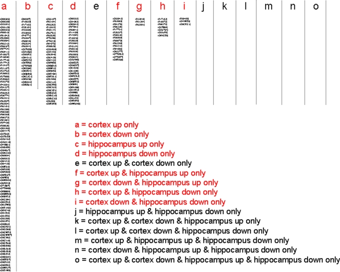
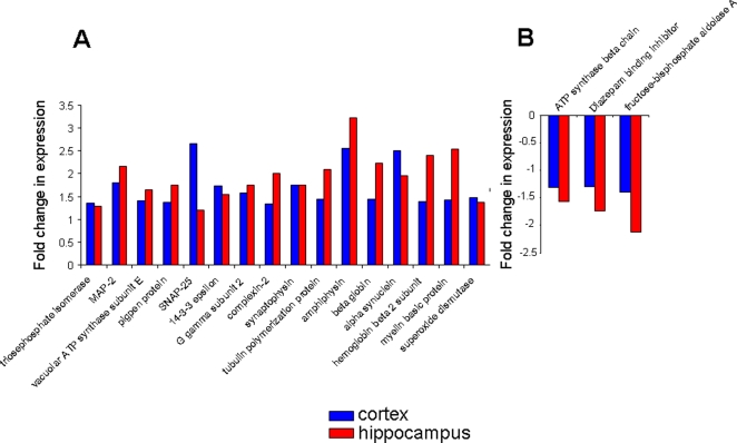
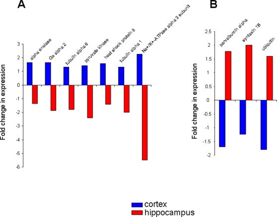
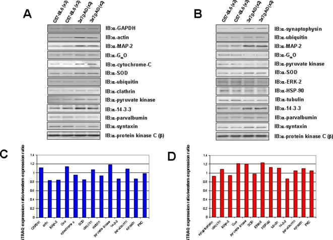
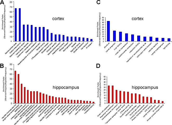
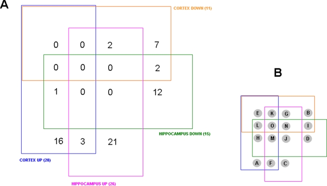
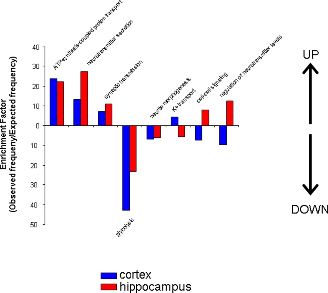
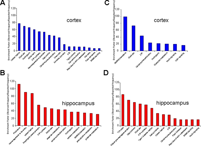
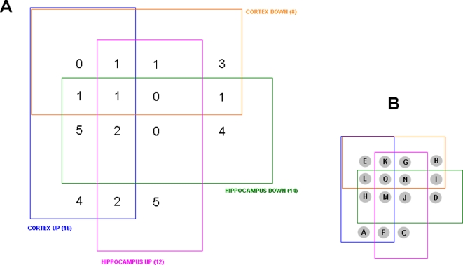
Alzheimer's disease (AD) is characterized by progressive cognitive impairment associated with accumulation of amyloid beta-peptide, synaptic degeneration and the death of neurons in the hippocampus, and temporal, parietal and frontal lobes of the cerebral cortex. Analysis of postmortem brain tissue from AD patients can provide information on molecular alterations present at the end of the disease process, but cannot discriminate between changes that are specifically involved in AD versus those that are simply a consequence of neuronal degeneration. Animal models of AD provide the opportunity to elucidate the molecular changes that occur in brain cells as the disease process is initiated and progresses. To this end, we used the 3xTgAD mouse model of AD to gain insight into the complex alterations in proteins that occur in the hippocampus and cortex in AD. The 3xTgAD mice express mutant presenilin-1, amyloid precursor protein and tau, and exhibit AD-like amyloid and tau pathology in the hippocampus and cortex, and associated cognitive impairment. Using the iTRAQ stable-isotope-based quantitative proteomic technique, we performed an in-depth proteomic analysis of hippocampal and cortical tissue from 16 month old 3xTgAD and non-transgenic control mice. We found that the most important groups of significantly altered proteins included those involved in synaptic plasticity, neurite outgrowth and microtubule dynamics. Our findings have elucidated some of the complex proteome changes that occur in a mouse model of AD, which could potentially illuminate novel therapeutic avenues for the treatment of AD and other neurodegenerative disorders.
阿尔茨海默病(AD)的特征是进行性认知障碍,与淀粉样β肽的积累、突触变性以及海马体、大脑皮层颞叶、顶叶和额叶中的神经元死亡有关。对AD患者死后脑组织的分析可以提供疾病过程末期存在的分子改变信息,但无法区分AD特异性涉及的变化与仅仅是神经元变性结果的变化。AD动物模型提供了一个机会,来阐明疾病过程开始和进展时脑细胞中发生的分子变化。为此,我们使用AD的3xTgAD小鼠模型来深入了解AD中海马体和皮层中发生的蛋白质复杂变化。3xTgAD小鼠表达突变的早老素-1、淀粉样前体蛋白和tau蛋白,并在海马体和皮层中表现出类似AD的淀粉样和tau病理,以及相关的认知障碍。使用基于iTRAQ稳定同位素的定量蛋白质组学技术,我们对16个月大的3xTgAD和非转基因对照小鼠的海马体和皮层组织进行了深入的蛋白质组分析。我们发现,显著改变的蛋白质中最重要的几组包括参与突触可塑性、神经突生长和微管动力学的蛋白质。我们的研究结果阐明了AD小鼠模型中发生的一些复杂蛋白质组变化,这可能为AD和其他神经退行性疾病的治疗开辟新的治疗途径。