Miao Haixi, Chen Lin, Riordan Sean M, Li Wenjun, Juarez Santiago, Crabb Andrea M, Lukas Thomas J, Du Pan, Lin Simon M, Wise Alexandria, Agapova Olga A, Yang Ping, Gu Charles C, Hernandez M Rosario
Department of Ophthalmology, Feinberg School of Medicine, Northwestern University, Chicago, Illinois, USA.
PLoS One. 2008 Aug 6;3(8):e2847. doi: 10.1371/journal.pone.0002847.
To determine whether optic nerve head (ONH) astrocytes, a key cellular component of glaucomatous neuropathy, exhibit differential gene expression in primary cultures of astrocytes from normal African American (AA) donors compared to astrocytes from normal Caucasian American (CA) donors.
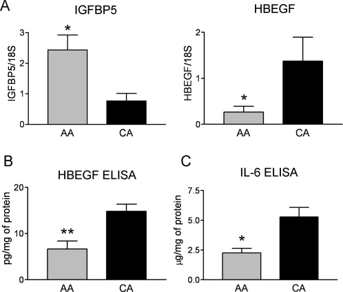
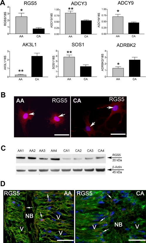
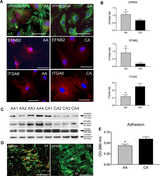
We used oligonucleotide Affymetrix microarray (HG U133A & HG U133A 2.0 chips) to compare gene expression levels in cultured ONH astrocytes from twelve CA and twelve AA normal age matched donor eyes. Chips were normalized with Robust Microarray Analysis (RMA) in R using Bioconductor. Significant differential gene expression levels were detected using mixed effects modeling and Statistical Analysis of Microarray (SAM). Functional analysis and Gene Ontology were used to classify differentially expressed genes. Differential gene expression was validated by quantitative real time RT-PCR. Protein levels were detected by Western blots and ELISA. Cell adhesion and migration assays tested physiological responses. Glutathione (GSH) assay detected levels of intracellular GSH.
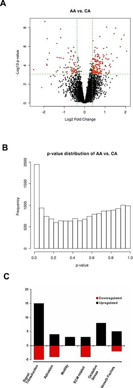
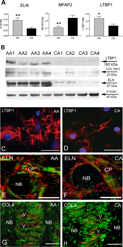
Multiple analyses selected 87 genes differentially expressed between normal AA and CA (P<0.01). The most relevant genes expressed in AA were categorized by function, including: signal transduction, response to stress, ECM genes, migration and cell adhesion.
These data show that normal astrocytes from AA and CA normal donors display distinct expression profiles that impact astrocyte functions in the ONH. Our data suggests that differences in gene expression in ONH astrocytes may be specific to the development and/or progression of glaucoma in AA.
确定青光眼性神经病变的关键细胞成分——视神经乳头(ONH)星形胶质细胞,与来自正常美国白种人(CA)供体的星形胶质细胞相比,在来自正常非洲裔美国人(AA)供体的星形胶质细胞原代培养物中是否表现出差异基因表达。
我们使用寡核苷酸Affymetrix微阵列(HG U133A和HG U133A 2.0芯片)来比较来自12名年龄匹配的CA正常供体眼睛和12名AA正常供体眼睛的培养ONH星形胶质细胞中的基因表达水平。芯片在R中使用Bioconductor通过稳健微阵列分析(RMA)进行标准化。使用混合效应模型和微阵列统计分析(SAM)检测显著的差异基因表达水平。功能分析和基因本体论用于对差异表达基因进行分类。通过定量实时RT-PCR验证差异基因表达。通过蛋白质印迹和ELISA检测蛋白质水平。细胞粘附和迁移试验测试生理反应。谷胱甘肽(GSH)试验检测细胞内GSH水平。
多项分析筛选出87个在正常AA和CA之间差异表达的基因(P<0.01)。AA中表达的最相关基因按功能分类,包括:信号转导、应激反应、细胞外基质基因、迁移和细胞粘附。
这些数据表明,来自AA和CA正常供体的正常星形胶质细胞表现出不同的表达谱,影响ONH中星形胶质细胞的功能。我们的数据表明,ONH星形胶质细胞中基因表达的差异可能是AA人群青光眼发生和/或进展所特有的。