Witschey Walter R T, Borthakur Arijitt, Elliott Mark A, Fenty Matthew, Sochor Matthew A, Wang Chenyang, Reddy Ravinder
Metabolic Magnetic Resonance Research and Computing Center, Department of Radiology, University of Pennsylvania, Philadelphia, Pennsylvania 19104-6100, USA.
J Magn Reson Imaging. 2008 Sep;28(3):744-54. doi: 10.1002/jmri.21444.
To develop a T1rho-prepared, balanced gradient echo (b-GRE) pulse sequence for rapid three-dimensional (3D) T1rho relaxation mapping within the time constraints of a clinical exam (<10 minutes), examine the effect of acquisition on the measured T1rho relaxation time and optimize 3D T1rho pulse sequences for the knee joint and spine.
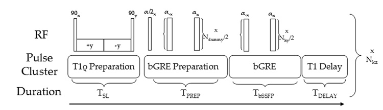
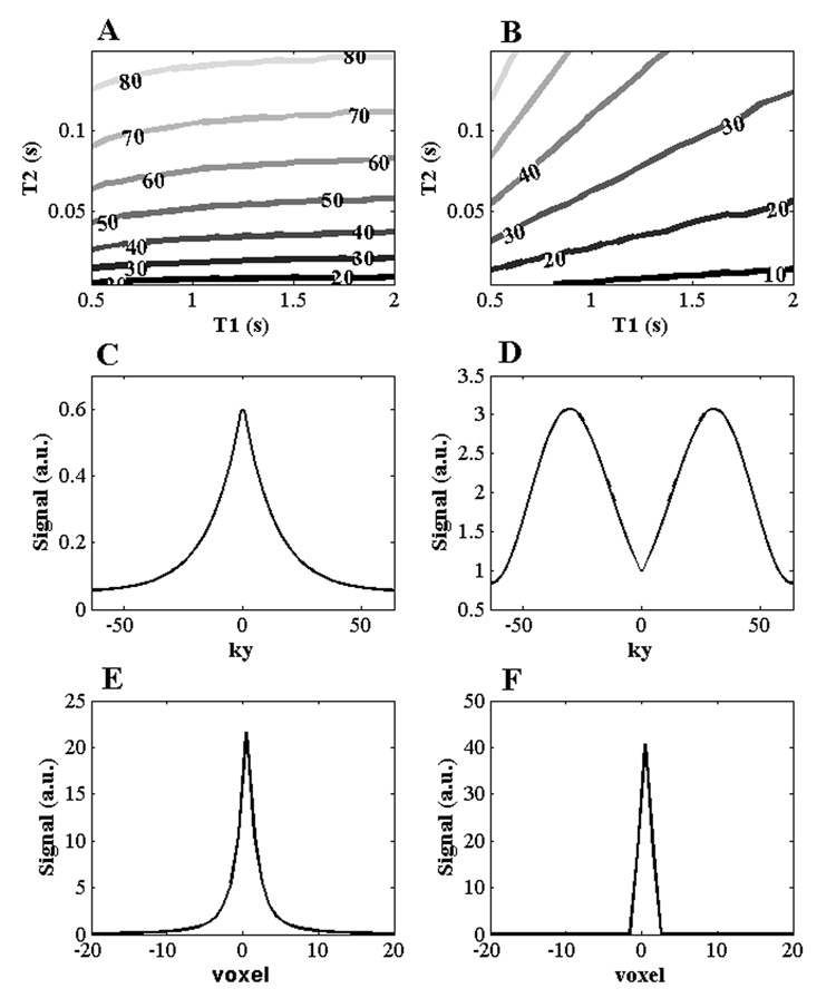
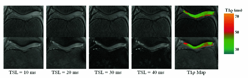
A pulse sequence consisting of inversion recovery-prepared, fat saturation, T1rho-preparation, and b-GRE image acquisition was used to obtain 3D volume coverage of the patellofemoral and tibiofemoral cartilage and lower lumbar spine. Multiple T1rho-weighted images at various contrast times (spin-lock pulse duration [TSL]) were used to construct a T1rho relaxation map in both phantoms and in the knee joint and spine in vivo. The transient signal decay during b-GRE image acquisition was corrected using a k-space filter. The T1rho-prepared b-GRE sequence was compared to a standard T1rho-prepared spin echo (SE) sequence and pulse sequence parameters were optimized numerically using the Bloch equations.
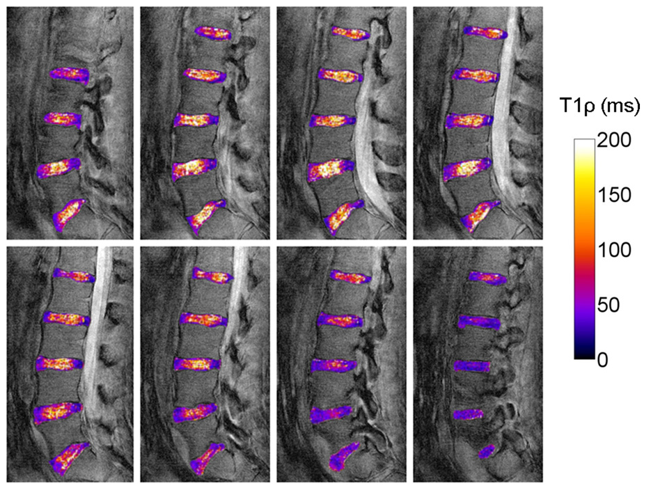
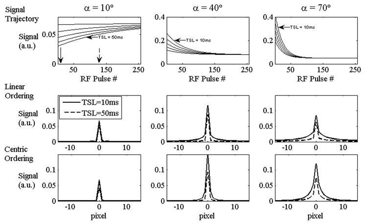
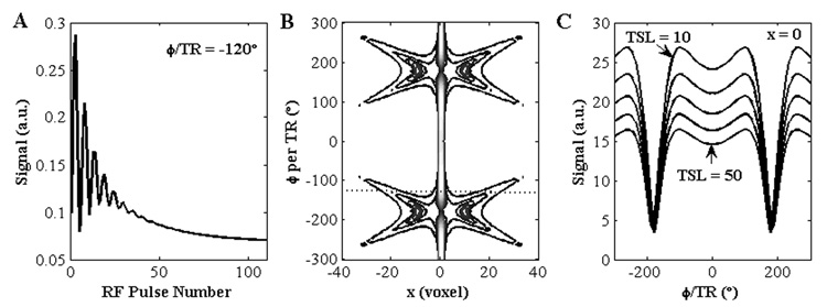
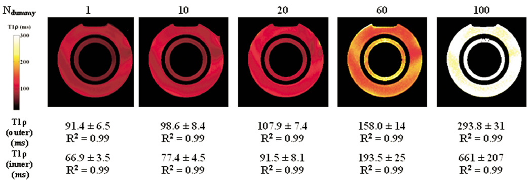
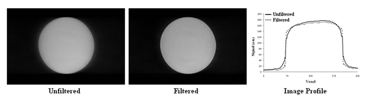
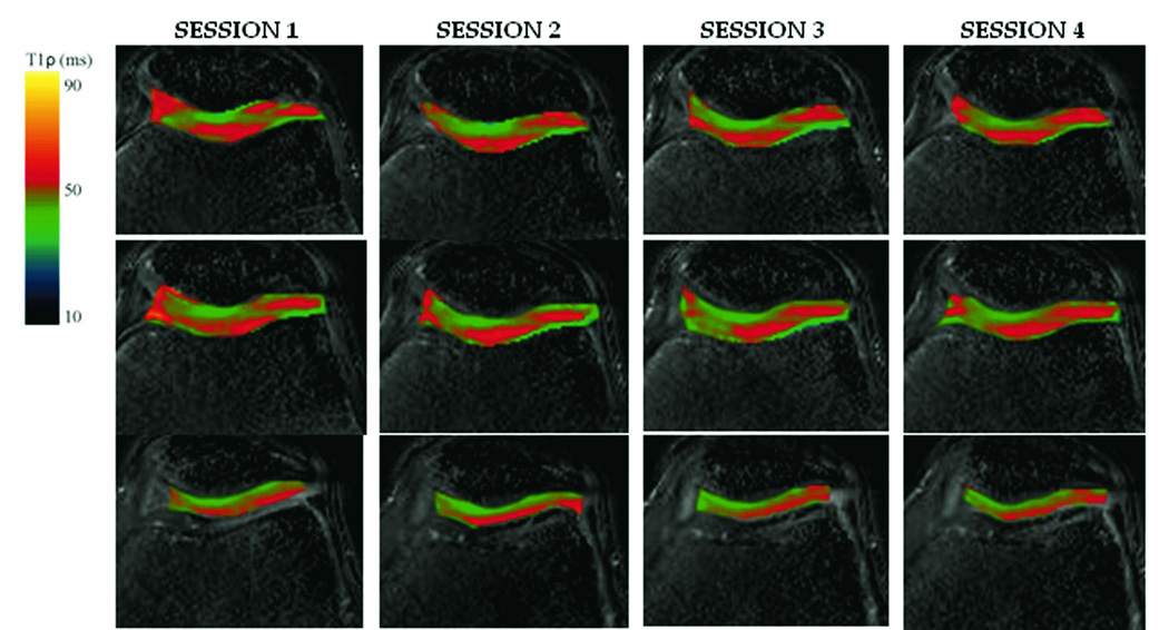
The b-GRE transient signal decay was found to depend on the initial T1rho-preparation and the corresponding T1rho map was altered by variations in the point spread function with TSL. In a two compartment phantom, the steady state response was found to elevate T1rho from 91.4+/-6.5 to 293.8+/-31 and 66.9+/-3.5 to 661+/-207 with no change in the goodness-of-fit parameter R2. Phase encoding along the longest cartilage dimension and a transient signal decay k-space filter retained T1rho contrast. Measurement of T1rho using the T1rho-prepared b-GRE sequence matches standard T1rho-prepared SE in the medial patellar and lateral patellar cartilage compartments. T1rho-preparedb-GRE T1rho was found to have low interscan variability between four separate scans. Mean patellar cartilage T1rho was elevated compared to femoral and tibial cartilage T1rho.
The T1rho-prepared b-GRE acquisition rapidly and reliably accelerates T1rho quantification of tissues offset partially by a TSL-dependent point spread function.
开发一种T1rho准备的平衡梯度回波(b-GRE)脉冲序列,用于在临床检查时间限制(<10分钟)内快速进行三维(3D)T1rho弛豫成像,研究采集对测量的T1rho弛豫时间的影响,并优化膝关节和脊柱的3D T1rho脉冲序列。
使用由反转恢复准备、脂肪饱和、T1rho准备和b-GRE图像采集组成的脉冲序列,获取髌股关节和胫股关节软骨以及下腰椎的3D体积覆盖图像。在不同对比时间(自旋锁定脉冲持续时间[TSL])下采集多个T1rho加权图像,用于构建体模以及体内膝关节和脊柱的T1rho弛豫图。使用k空间滤波器校正b-GRE图像采集期间的瞬态信号衰减。将T1rho准备的b-GRE序列与标准的T1rho准备的自旋回波(SE)序列进行比较,并使用布洛赫方程对脉冲序列参数进行数值优化。
发现b-GRE瞬态信号衰减取决于初始T1rho准备,并且相应的T1rho图会因TSL导致的点扩散函数变化而改变。在双室体模中,发现稳态响应使T1rho从91.4±6.5升高到293.8±31,从66.9±3.5升高到661±207,而拟合优度参数R2没有变化。沿着最长软骨维度进行相位编码和瞬态信号衰减k空间滤波器保留了T1rho对比度。使用T1rho准备的b-GRE序列测量的T1rho与标准的T1rho准备的SE在髌内侧和髌外侧软骨区域匹配。发现T1rho准备的b-GRE T1rho在四次单独扫描之间具有较低的扫描间变异性。与股骨和胫骨软骨的T1rho相比,髌软骨的平均T1rho升高。
T1rho准备的b-GRE采集快速且可靠地加速了T1rho组织定量,部分被TSL依赖的点扩散函数所抵消。